Koala (Phascolarctos cinereus)
Family Phascolarctidae
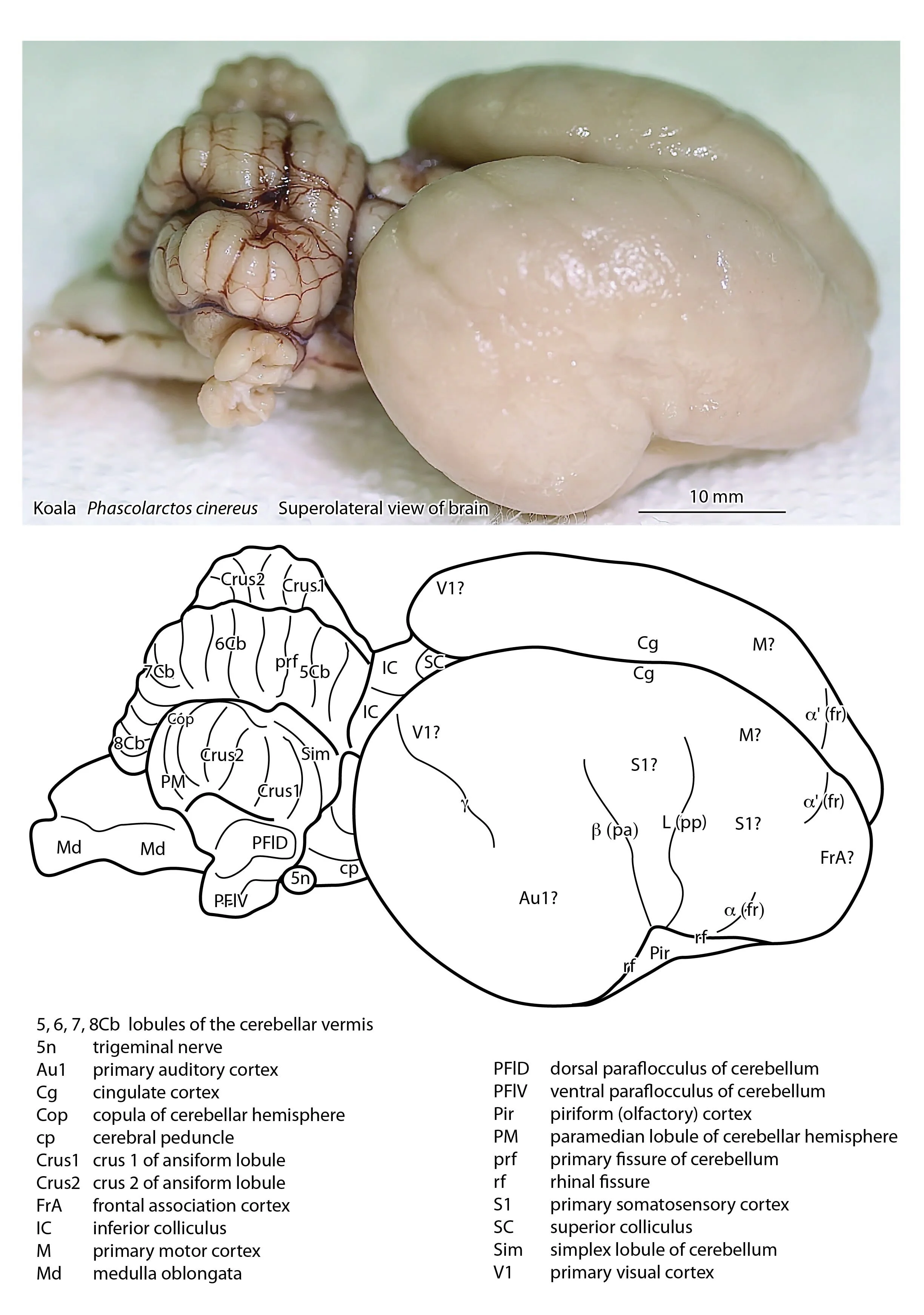
The brain of the koala is about 20 ml in volume. The cerebral hemispheres are relatively smooth (lissencephalic) and reduced in size compared with other diprotodontids (Haight and Nelson, 1987), such that the superior and inferior colliculi, pineal gland, and even the roof of the third ventricle are exposed. This reduction in cortical size (and therefore cerebral metabolism) may be an adaptation to the poor quality of their diet. The functional areas of the cerebral cortex have not been assessed physiologically, so the labelling of S1, V1 and Au1 in the accompanying illustrations is putative based on cellular patterns in neuronal (Nissl) stained specimens from the Nelson collection. The cerebellum is foliated, but the cerebellar hemispheres are not as elaborate as in other diprotodontids, reflecting poor development of the cerebro(ponto)cerebellar circuitry.
Reference
Haight JR, Nelsons JE (1987) A brain that doesn’t fit its skull: a comparative study of the brain and endocranium of the koala Phascolarctos cinereus (Marsupialia: Phascolarctidae). In Possums and Opossums: Studies in Evolution (ed. M. Archer). Sydney: Surrey Beatty and Sons. pp. 331-352.