Atlas of the Brain of a 4 week Human Embryo, Carnegie #836
Introduction
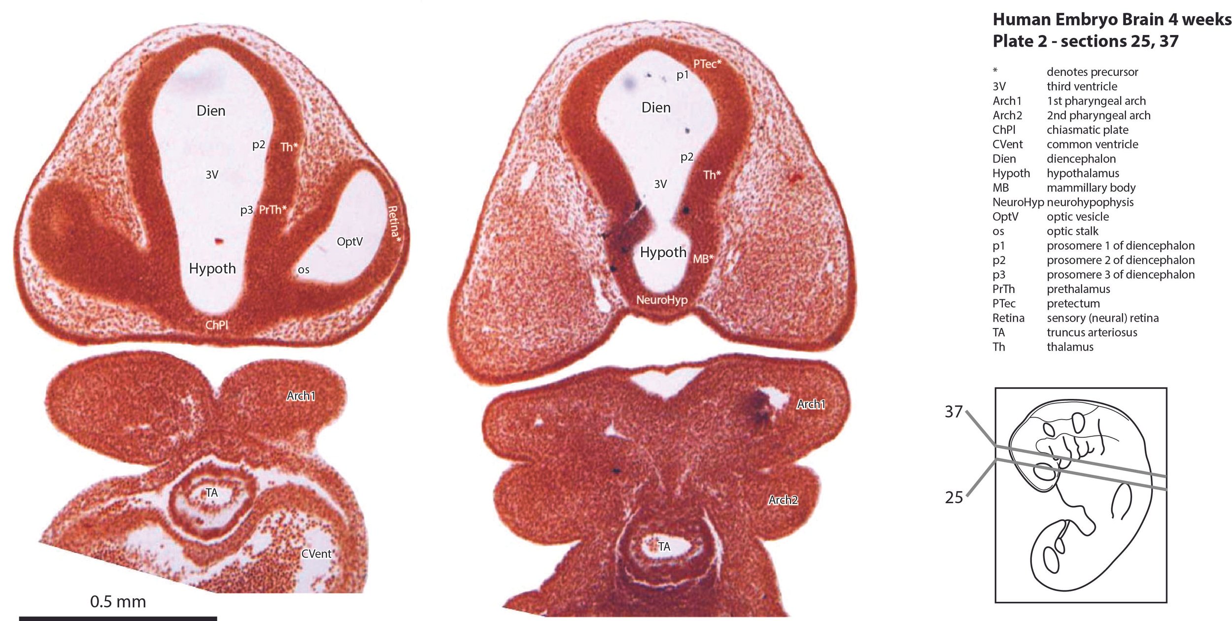
This specimen is at a stage (Stage 13) shortly after neural tube closure, when the forebrain vesicle has developed, but lateral evagination of the telencephalic vesicles has not yet occurred. Division into hypothalamic, prosomeric, mesencephalic, isthmic and rhombencephalic segments is evident. The brain tissue is made up of proliferative compartments (neuroepithelium), with negligible populations of (post-mitotic) neurons confined to the rhombencephalon.
Methods
The specimen illustrated here is Carnegie #836, held at the National Museum for Health and Medicine (NMHM) in Silver Springs, Maryland. Likely crown-rump length is 4 mm. Estimated post-conception age is approximately 28 to 30 days.
The specimen was acquired in 1914, fixed in acetic acid, embedded in paraffin, sectioned transversely at a thickness of 15 µm, and the sections stained with alum cochineal (O’Rahilly and Müller, 1987).
Raw images were collected by Ken Ashwell at the NMHM in 2014, using a Nikon E800 microscope with a Ludl Biopoint automated stage and a MAC 2000 controller. Photomicrographs were taken with Micropublisher V5, with ImagePro Plus software from Media Cybernetics.
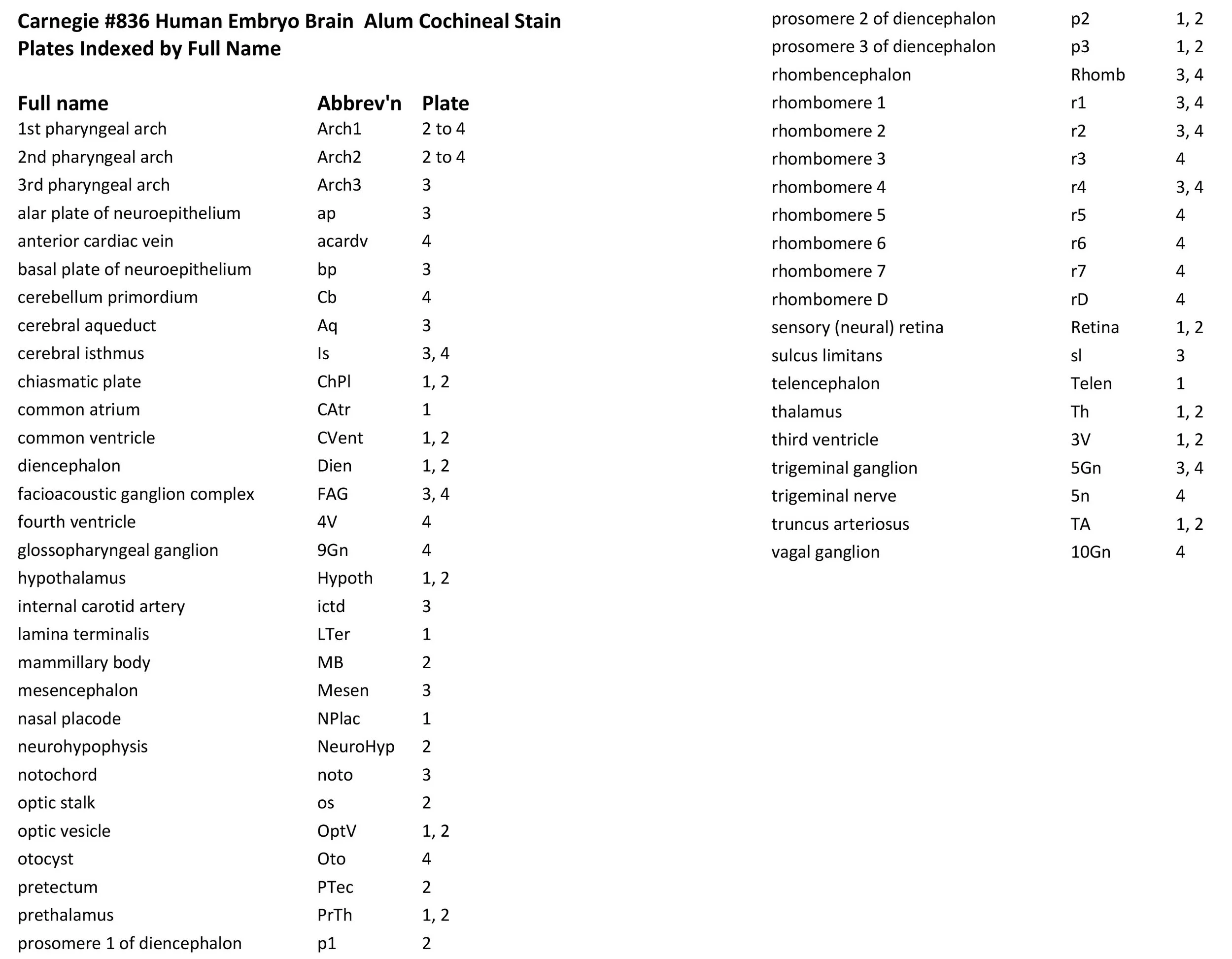
All images were calibrated by photographing a scale bar at the same magnification. Images were photomontaged and optimised for contrast using Adobe Photoshop 2023 before being placed in Adobe Illustrator 2023 for delineation and labelling. Developmental regions (i.e. neuroepithelium) destined to give rise to adult structures have been denoted by the adult structure’s name with an asterisk (e.g. Th* denotes the developmental field of the dorsal thalamus).
A line diagram of a stage 13 human embryo (O’Rahilly and Müller, 2006) has been used for the finder illustration indicating section position.
Notes on the specimen
General observations
Neural tube closure is complete (ORahilly and Müller, 2006), but there is no choroid plexus present. The major sensory ganglia have appeared (see below) and the first neurons are being generated for the rhombencephalon.
Central nervous system
The telencephalon is still a midline structure, because evagination of the telencephalic vesicles has not yet occurred. A chiasmatic plate, optic stalk, neurohypophysis and mammillary region are evident within the hypothalamus (see variously plates 1 and 2). It is possible to discern three prosomeric regions within the diencephalon (p1 -> pretectum, p2 -> dorsal thalamus, p3 -> prethalamus, from caudal to rostral; see plates 1, 2). The mesencephalon has developed a marginal layer, predominantly composed of longitudinally running axons. The rhombencephalon show a distinct rhombomeric organisation (r1 to r7, rD; see plate 4) and a thin marginal layer where some of the first generated neurons will come to rest. The cerebellum makes its first appearance as a thickening of the alar plate of rhombomere 1 (plate 4).
Sense organs and peripheral nervous system
The nasal placodes are evident (NPlac in plate 1), but there has not been any invagination to form a nasal cavity. The optic vesicle (OptV in plates 1, 2) has extended laterally from the forebrain vesicle, but has not yet formed an optic cup. Putative sensory retina tissue has been labelled (Retina in plates 1, 2). The trigeminal ganglion lies alongside rhombomere 2 (please see 5Gn in plate 4) and extends a trigeminal nerve (5n) into the rhombencephalon. The otic vesicle (Oto in plate 4) has separated from the ectoderm to lie alongside rhombomere 5, but is still a simple ellipsoidal shape with no diverticula. The facioacoustic ganglion complex (FAG in plate 4) is a single mass alongside rhombomere 4, i.e. distinct geniculate, vestibular and cochlear components have yet to appear. Glossopharyngeal and vagal ganglia can be seen (9Gn and 10Gn alongside rhombomeres 6 and 7, respectively, in plate 4).
Acknowledgements
I would like to thank Dr Elizabeth Lockett of the NMHM for access to the collection and for all her kind help during the work.
References
O’Rahilly R, Müller F (1987) Developmental stages in human embryos. Carnegie Institution of Washington 63T.
O’Rahilly R, Müller F (2006) The Embryonic Human Brain. An Atlas of Developmental Stages. Wiley, Hoboken.