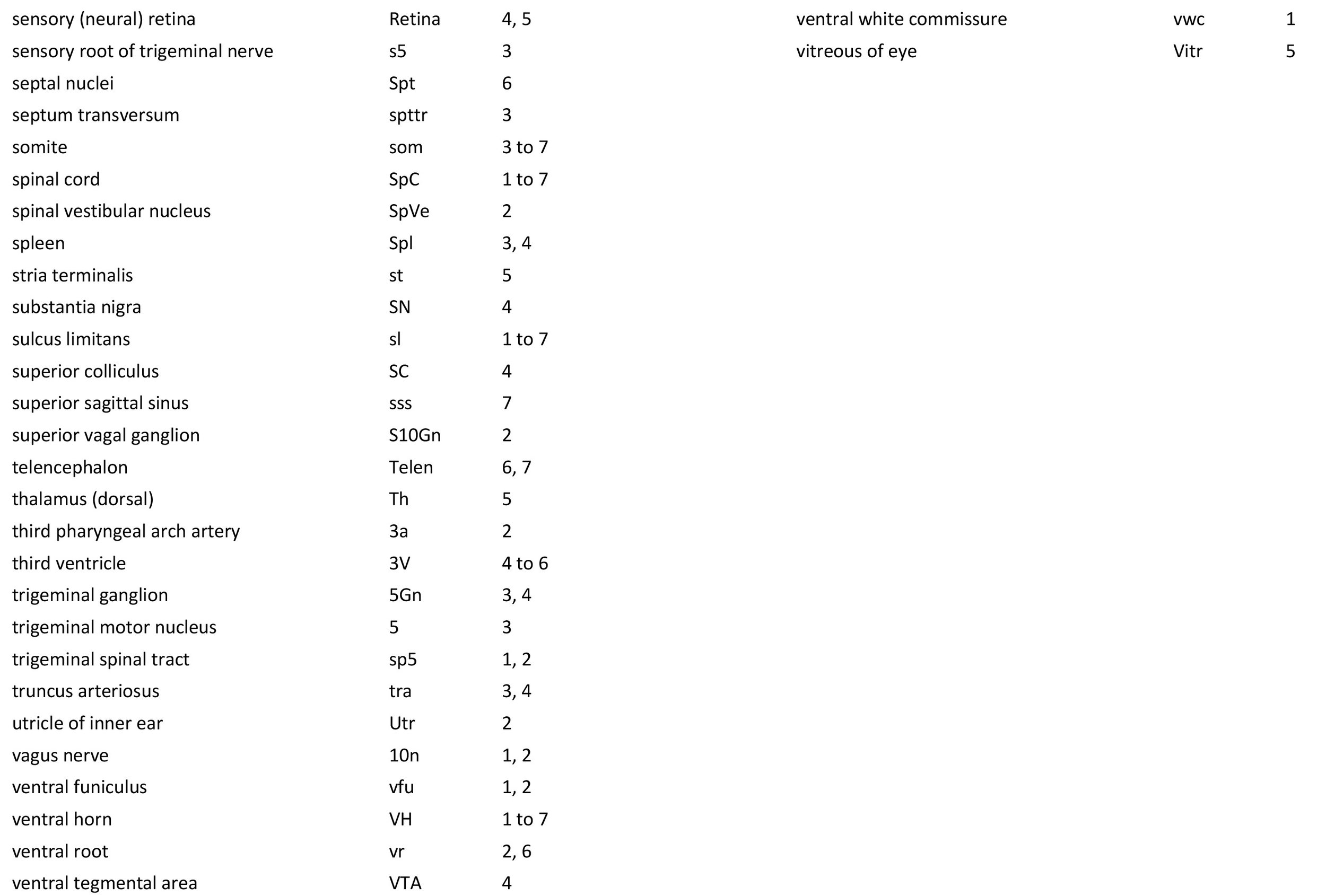
Atlas of an E13 Rat Embryo
Introduction
This series of images illustrates the state of development of major organ systems in the laboratory rat at the end of the embryonic period. The embryo was approximately 6.5 mm in greatest length (GL) when fresh or approximately 4.5 mm GL after fixation and dehydration.
Methods
All procedures were carried out in accordance with accepted ethical principles for animal research and were approved by the relevant Animal Ethics Committee of the University of New South Wales.
Rat embryos and fetuses were obtained from timed matings, where the first 24 hours after conception was designated as E0. The specimen illustrated here was fixed by immersion in Bouin’s fixative for 12 hours, dehydrated in ascending ethanol concentrations, cleared in histolene and subsequently embedded in paraffin. Sections were cut frontally at 7 µm thickness and stained with hematoxylin and eosin before coverslipping.
General observations
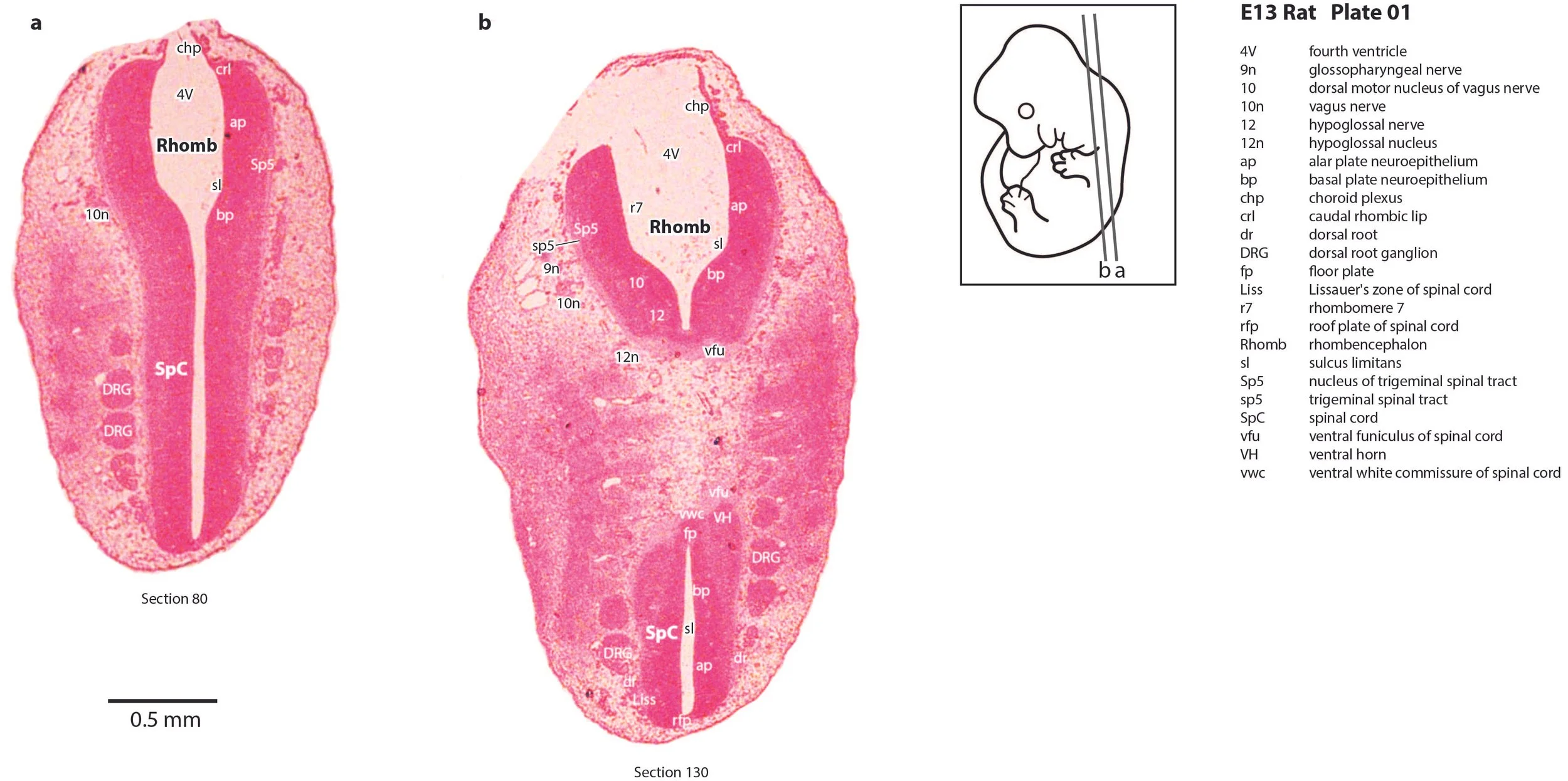
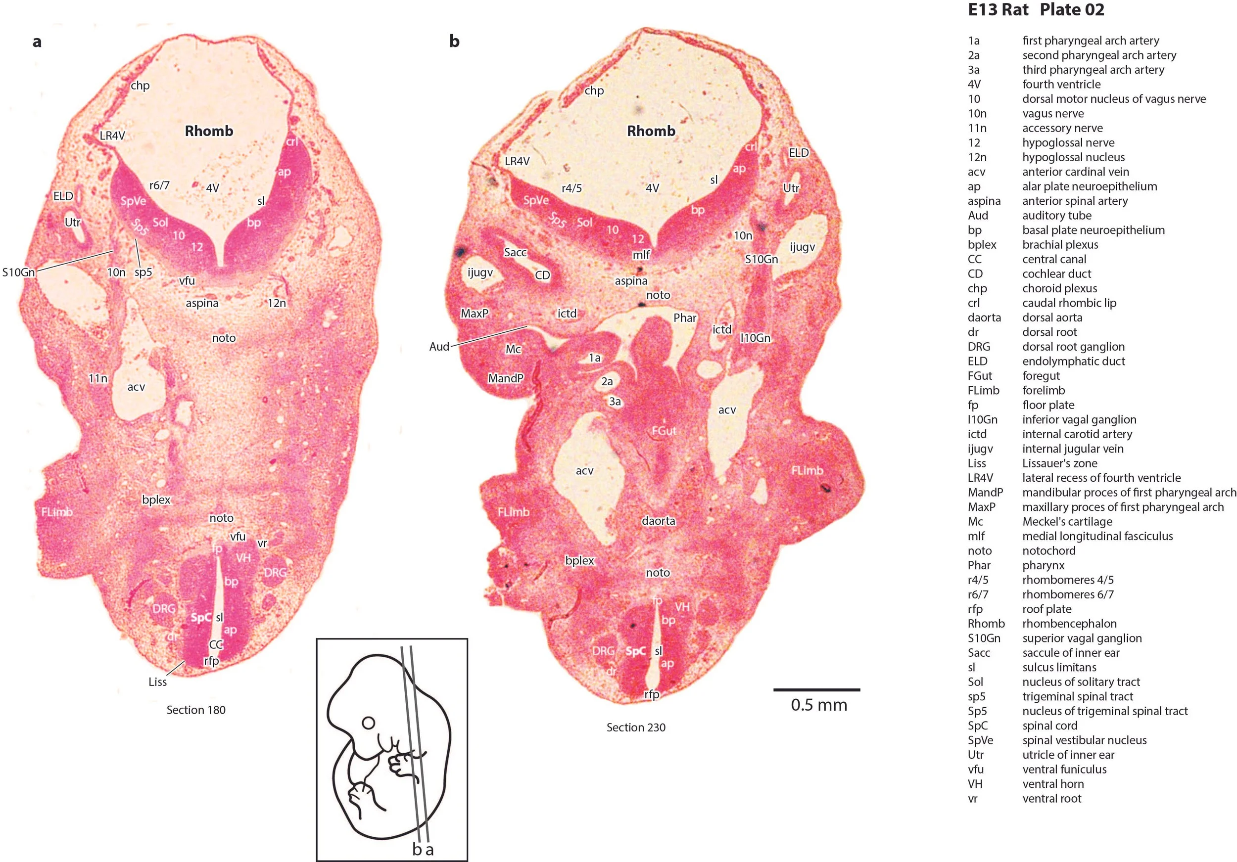
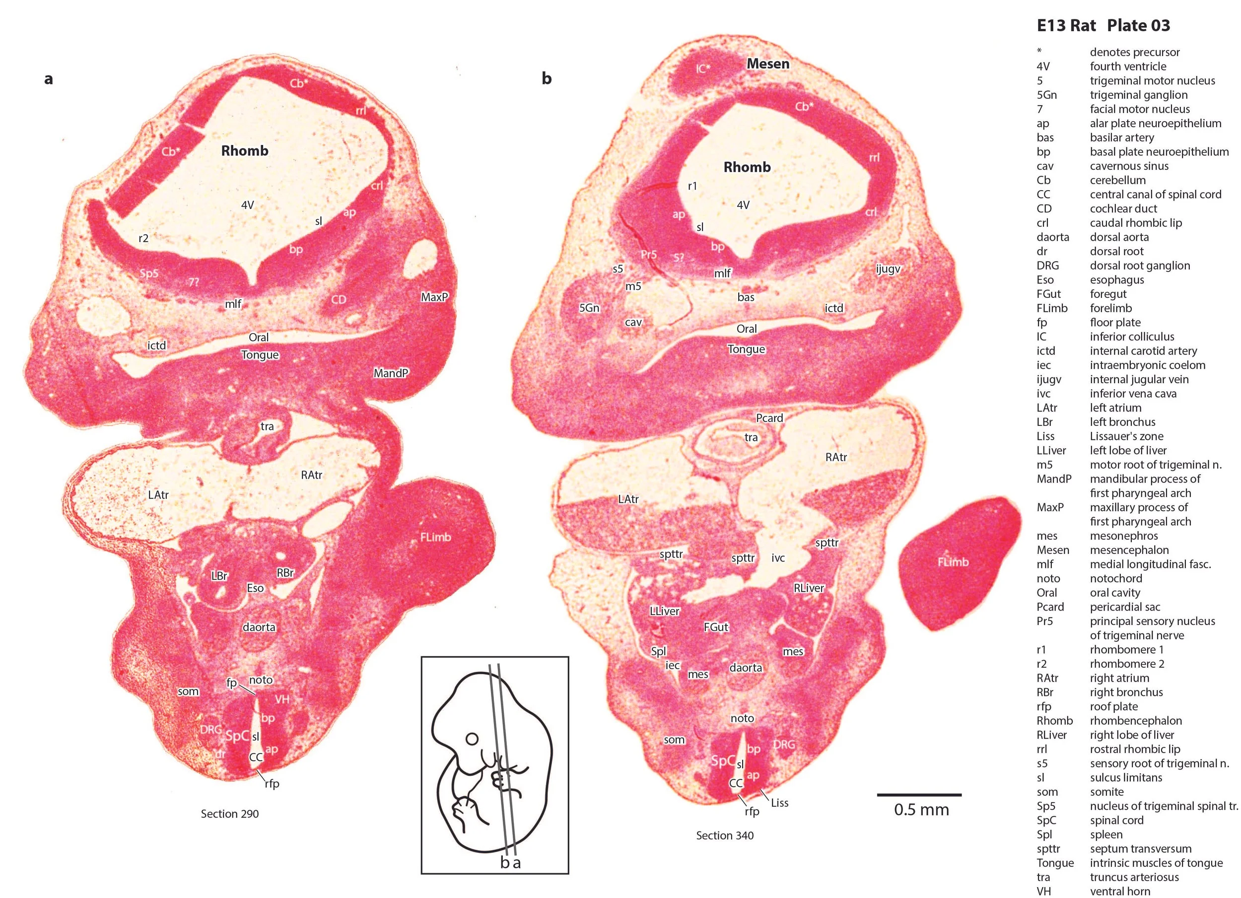
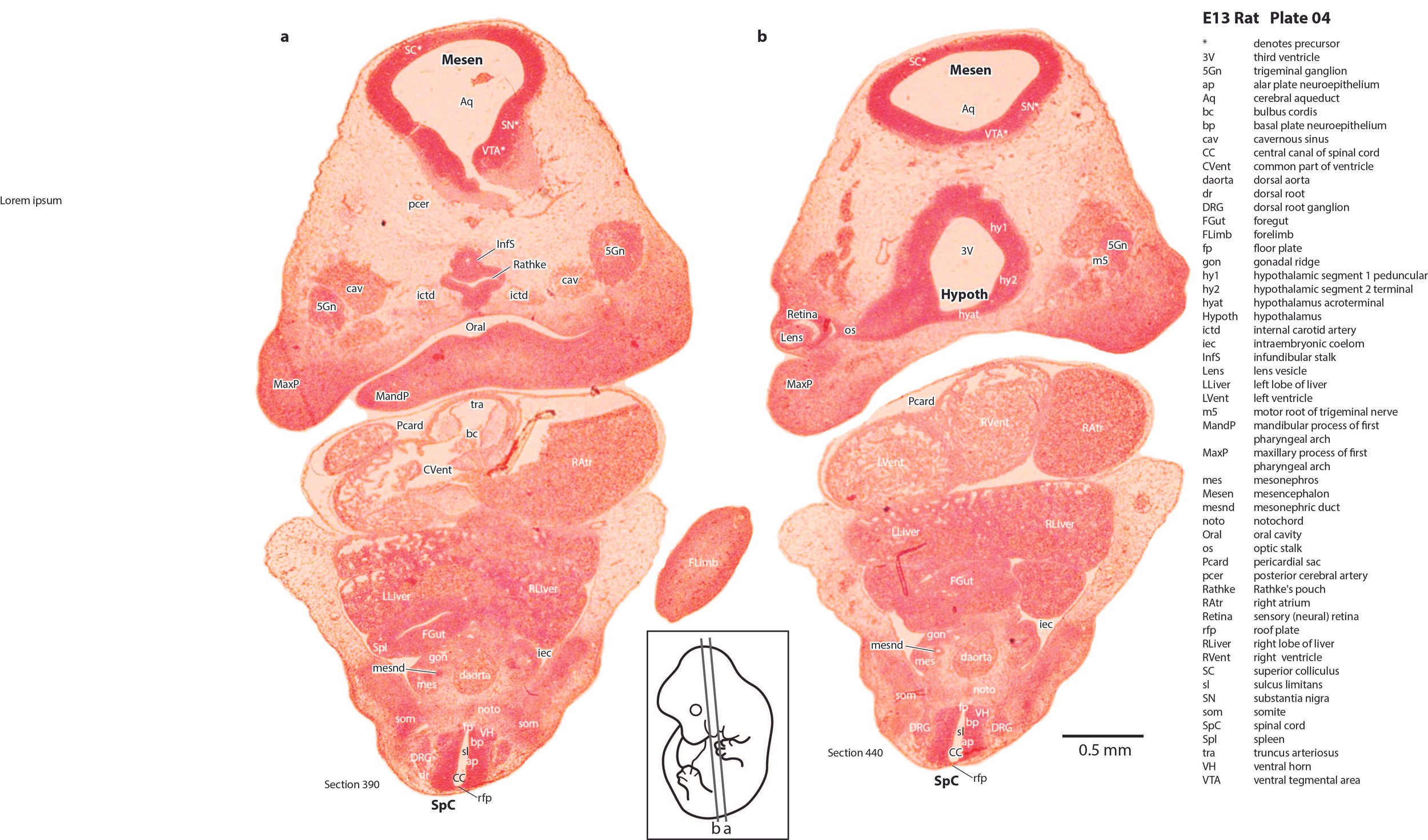
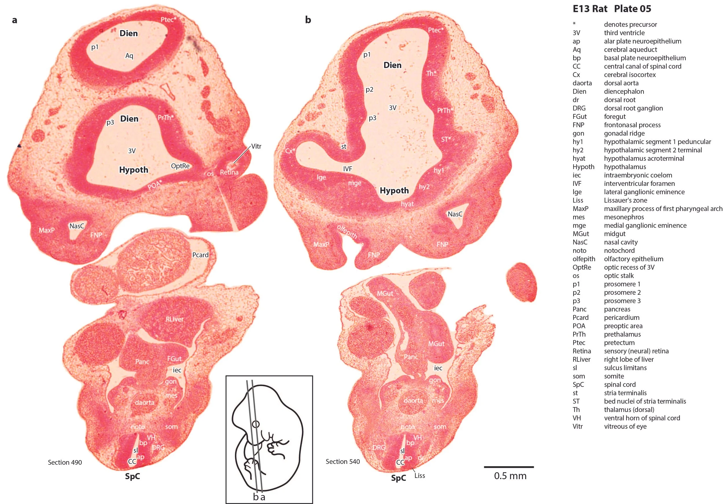
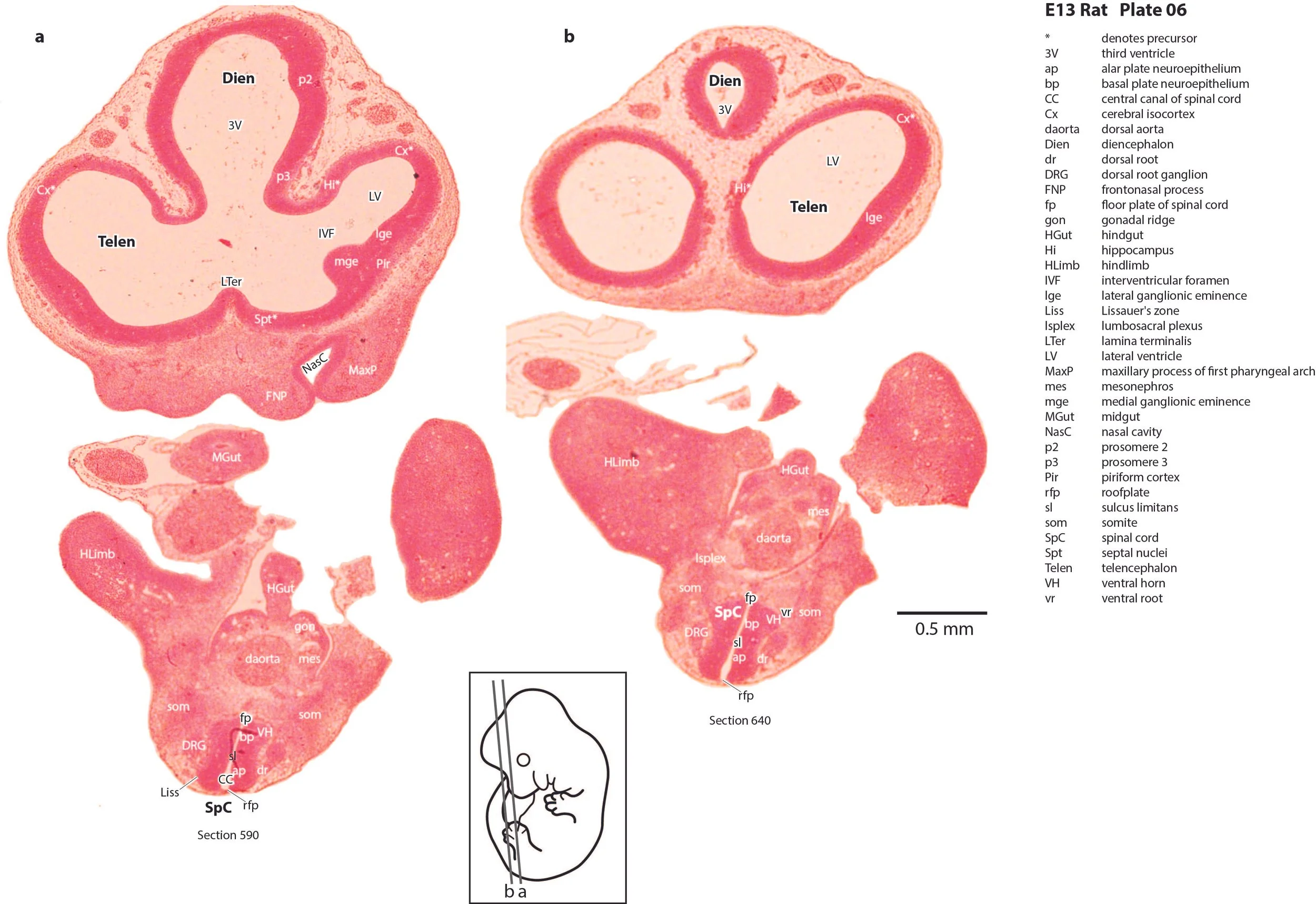
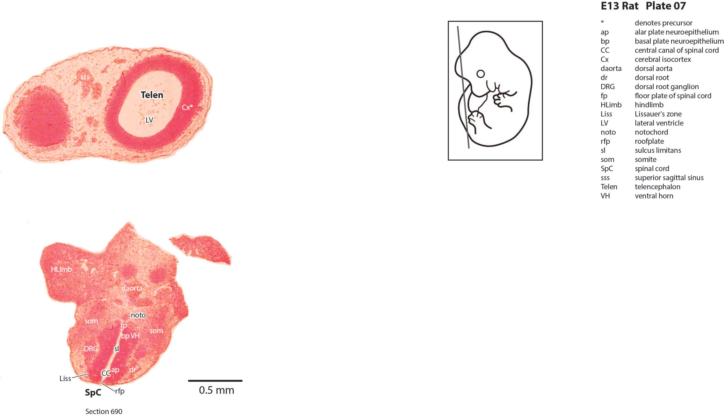
The specimen is at the late embryonic stage, meaning that some distinctly embryonic structures (e.g. pharyngeal arch arteries, mesonephros, truncus arteriosus) are still visible. The neural tube has elaborated telencephalic vesicles and optic cups, but major neurogenesis has yet to commence.
The ventricle has begun separation into distinct left and right parts, but still has a common region (CVent in Plate 4) leading to the bulbus cordis and truncus arteriosus.
Olfactory apparatus
The nasal cavity has formed between the frontonasal and maxillary processes, but the olfactory epithelium (olfepith in Plate 5) is still primitive. A rudimentary piriform cortex (Pir in Plate 6) is visible.
Telencephalon
The cerebral cortex (Cx – Plates 5 to 7) or pallium is relatively thin at this developmental stage, with few postmitotic neurons evident. The hippocampus (Hi – Plate 6) is also very poorly differentiated. Lateral and medial ganglionic eminences (lge, mge in Plates 5 and 6) have appeared, but only a few striatal and pallidal neurons have been generated. The septal region (Spt in Plate 6) is composed solely of neuroepithelium.
Diencephalon
Prosomeres 1 to 3 (Puelles et al. 2012b) are visible in the diencephalon (see Plates 5 and 6). but no postmitotic neurons are visible.
Hypothalamus
Developmental subdivisions of the hypothalamus (peduncular, tuberal and acroterminal hypothalamus; hy1, hy2, hyat, respectively) described by Puelles and co-workers (Puelles et al. 2012a) have been identified here (Plates 4 and 5).
Cerebellum
There is a rostral rhombic lip (rrl in Plate 3) forming the roof of the fourth ventricle, but cerebellar neurogenesis has not yet initiated.
Brainstem
Alar and basal plates are visible separated by the sulcus limitans. Some major motor and sensory nuclei have begun to settle adjacent to the pial membrane.
References
Puelles L, Martinez-de-la-Torre M, Bardet S and Rubenstein JLR (2012a) Hypothalamus. In The Mouse Nervous System (Eds C Watson, G Paxinos and L Puelles) pp. 221–312. Elsevier, San Diego.
Puelles L, Martinez-de-la-Torre M, Ferran J-L and Watson CRR (2012b) Diencephalon. In The Mouse Nervous System (Eds C Watson, G Paxinos and L Puelles) pp. 313–336. Elsevier, San Diego.