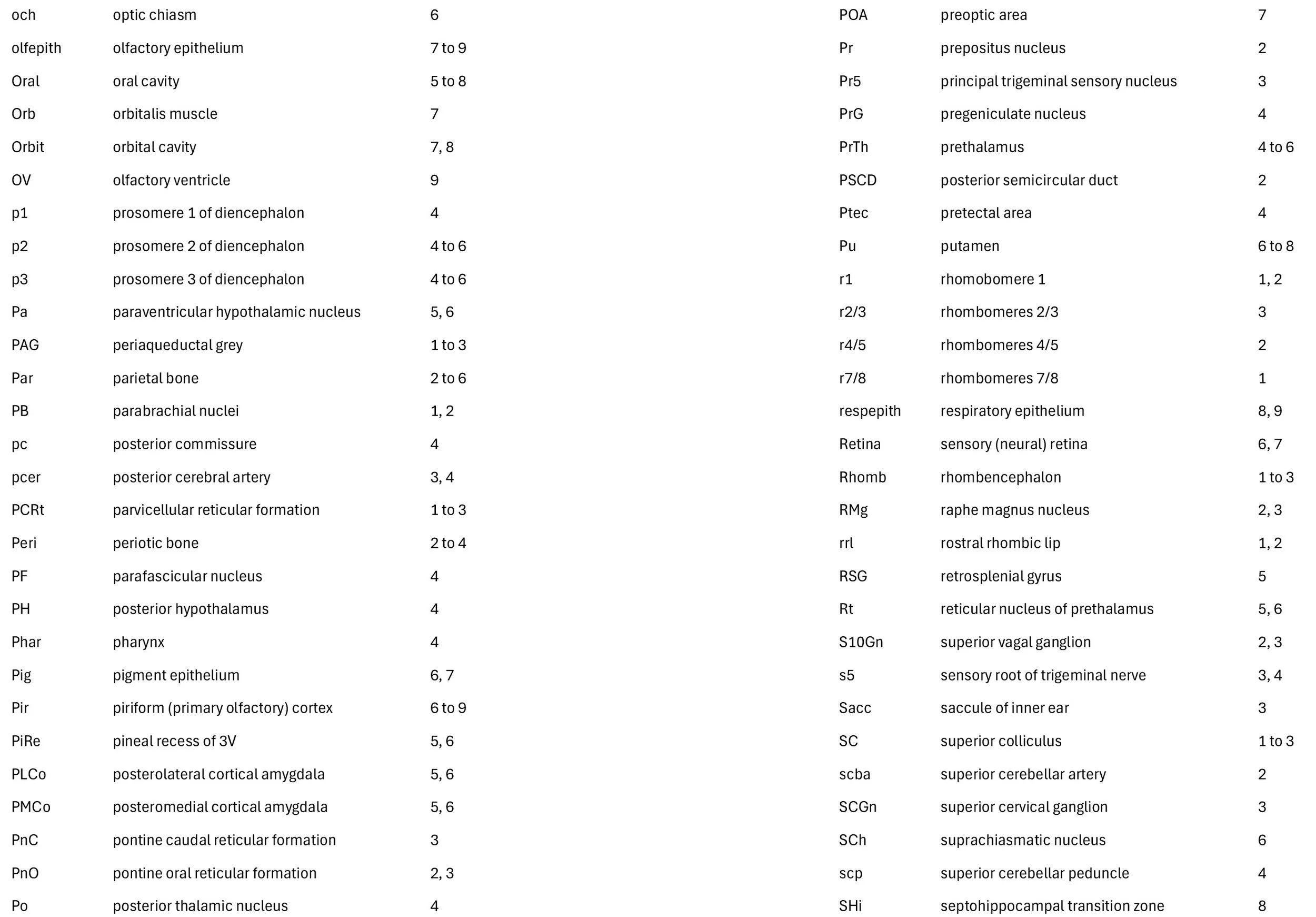
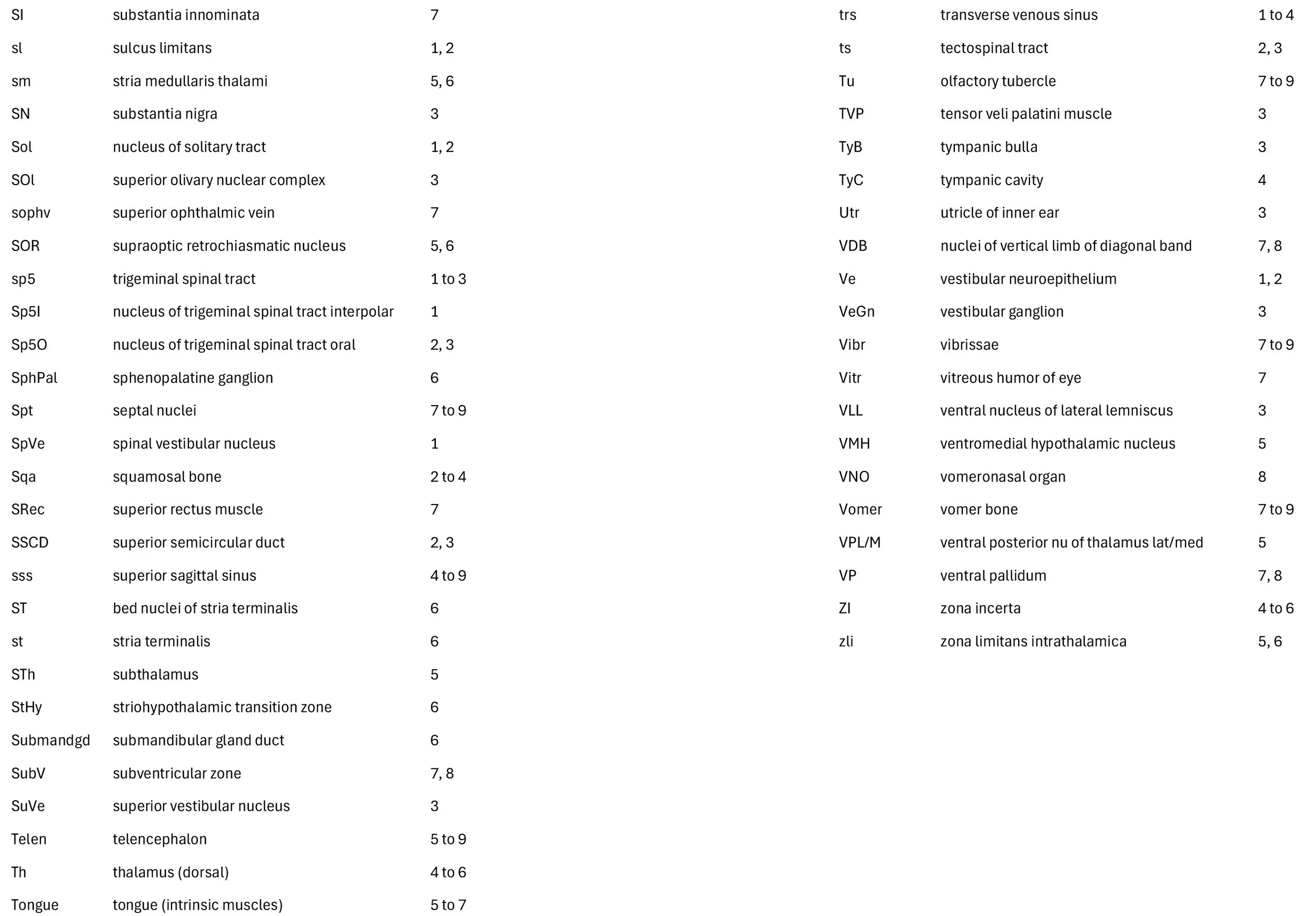
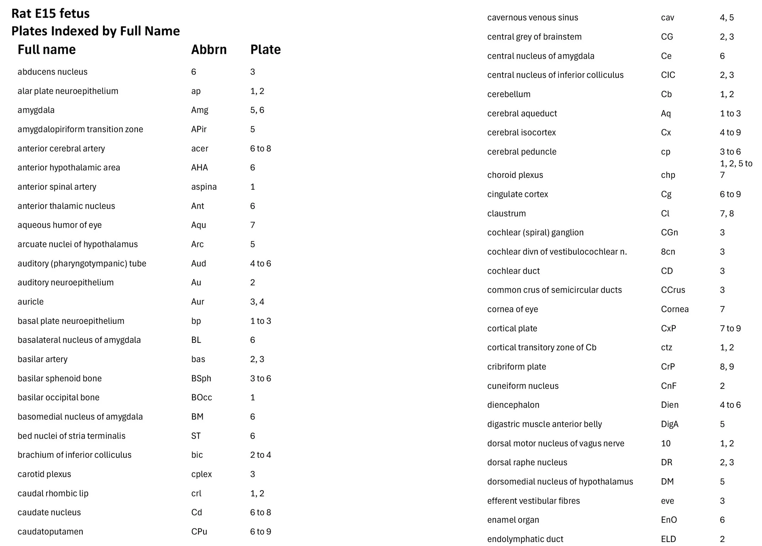
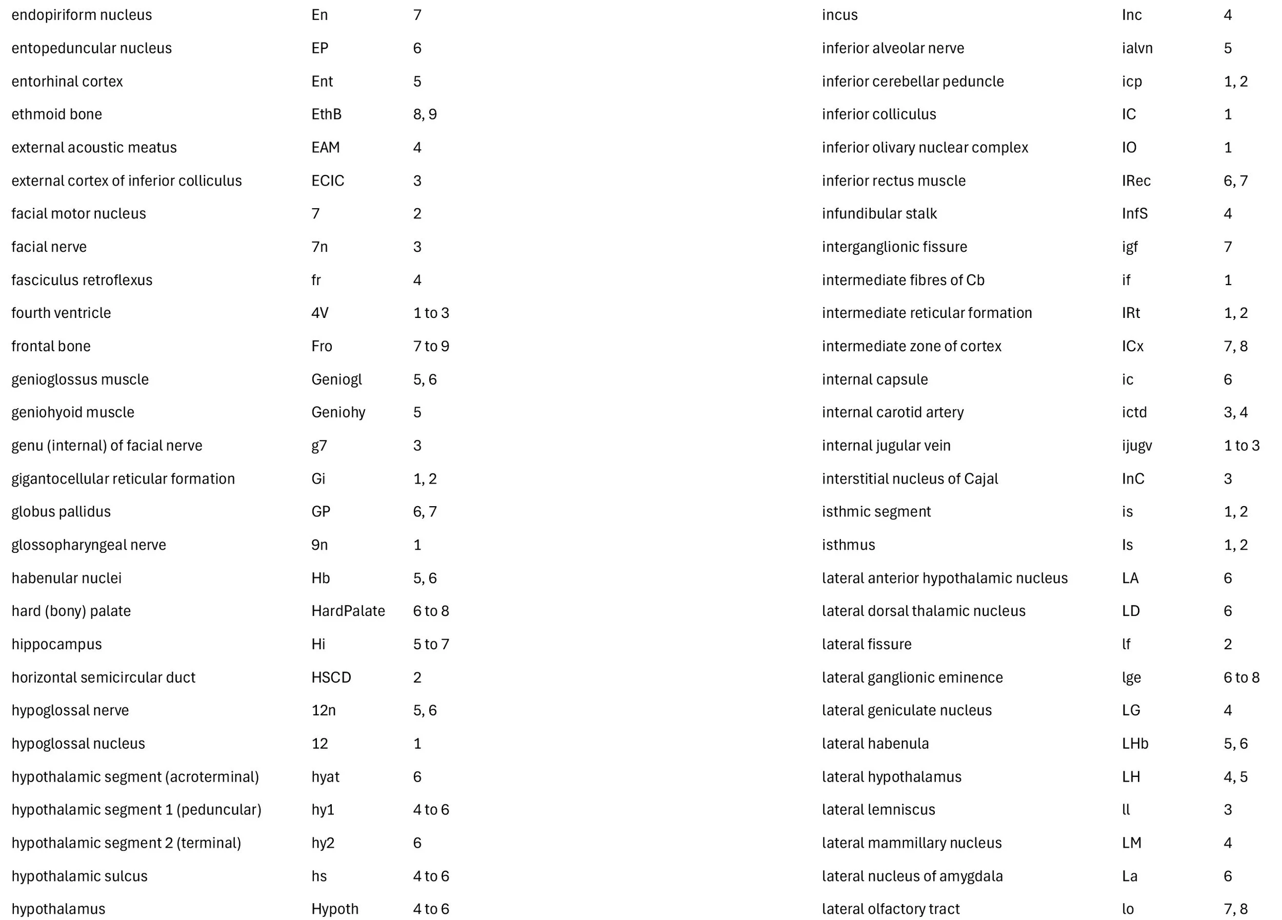
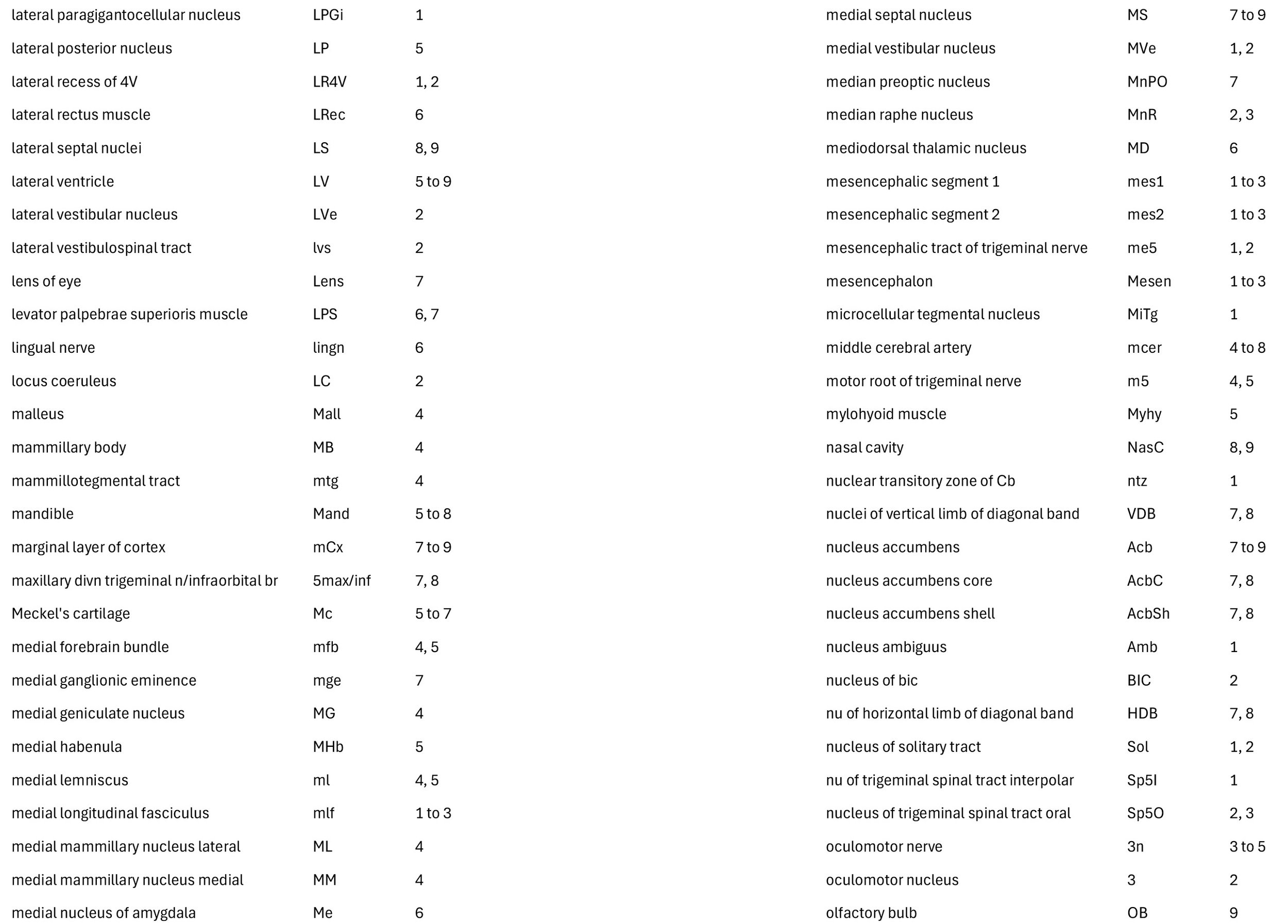
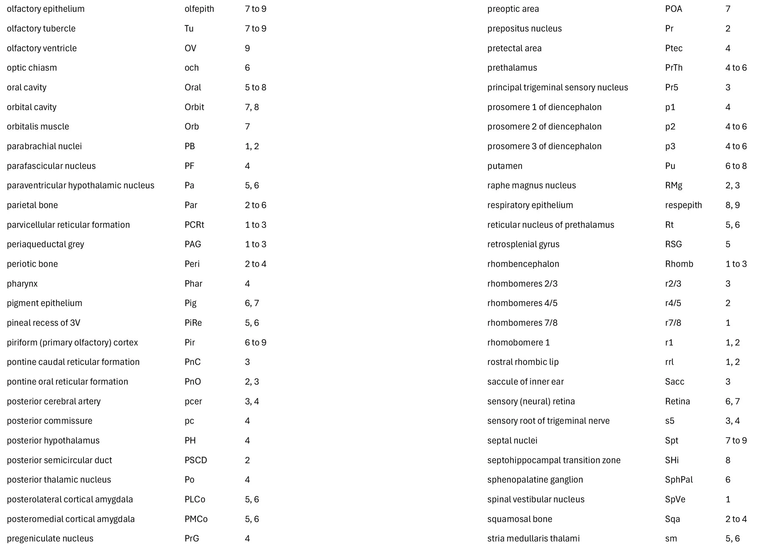
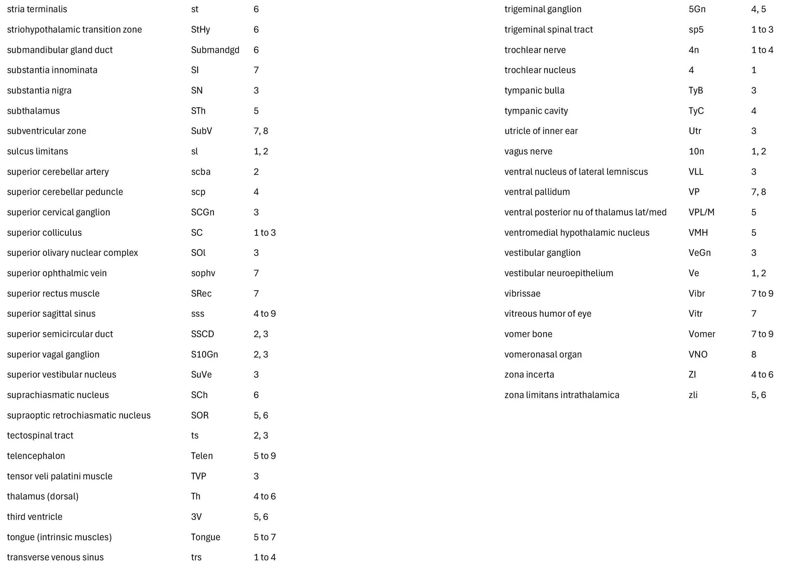
Atlas of the Brain of an E15 Rat Fetus
Introduction
This series of images illustrates the state of development of the brain in a laboratory rat at the beginning of the fetal period.
The fetus was approximately 8.5 mm in greatest length (GL) when fresh or approximately 6.0 mm GL (4 mm head width) after fixation and dehydration.
Methods
All procedures were carried out in accordance with accepted ethical principles for animal research and were approved by the relevant Animal Ethics Committee of the University of New South Wales.
Rat embryos and fetuses were obtained from timed matings, where the first 24 hours after conception was designated as E0. The specimen illustrated here was fixed by immersion in Bouin’s fixative for 18 hours, dehydrated in ascending ethanol concentrations, cleared in histolene and subsequently embedded in paraffin. Sections were cut frontally at 7 µm thickness and stained with hematoxylin and eosin before coverslipping.
General observations
The specimen is at the early fetal stage, meaning that all organ systems are present and distinctly embryonic structures (e.g. pharyngeal arch arteries, mesonephros, truncus arteriosus) are transforming into adult structures. In the brain, major neurogenesis is underway in the walls of the brain vesicles, producing the macroneurons which provide the long connections within the central nervous system.
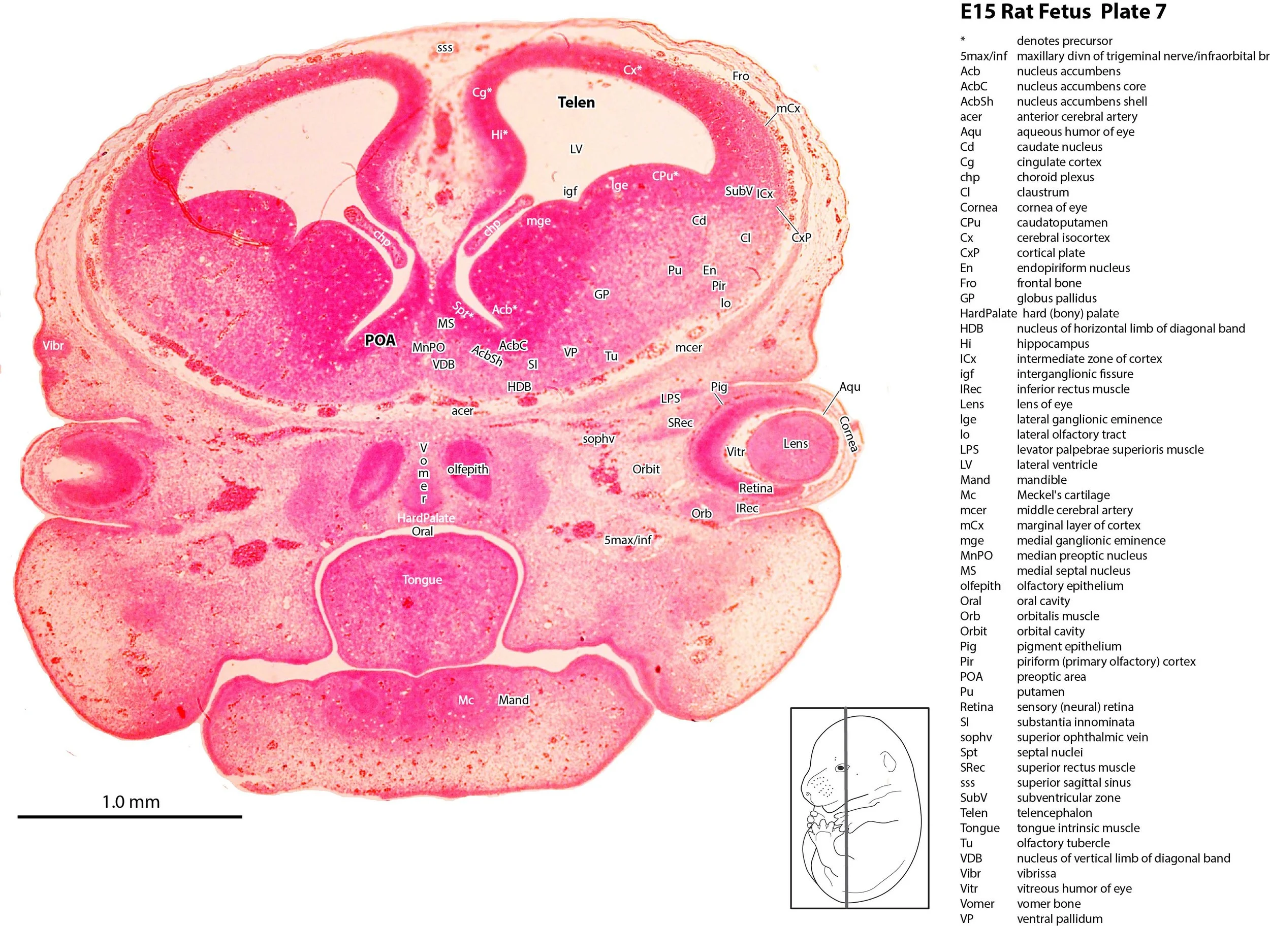
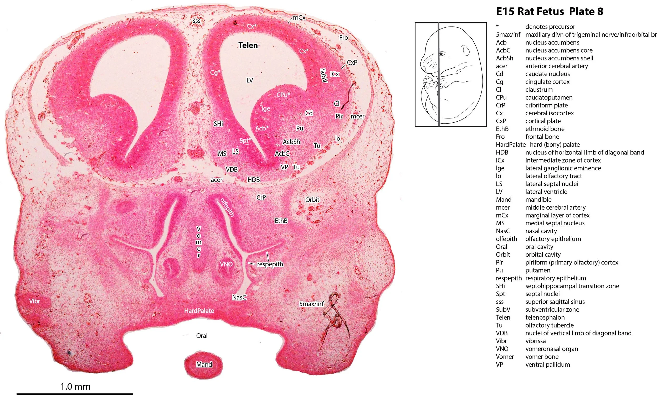
Olfactory apparatus
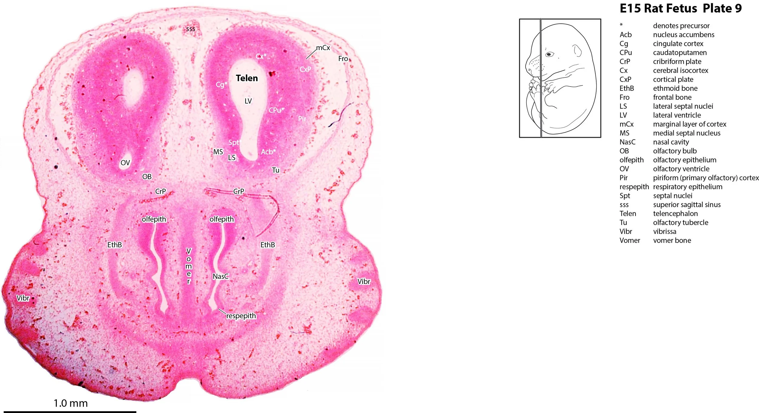
The nasal cavity has formed and elaboration of the nasal turbinates is beginning. Olfactory and respiratory epithelium can be distinguished (Plates 7 to 9).
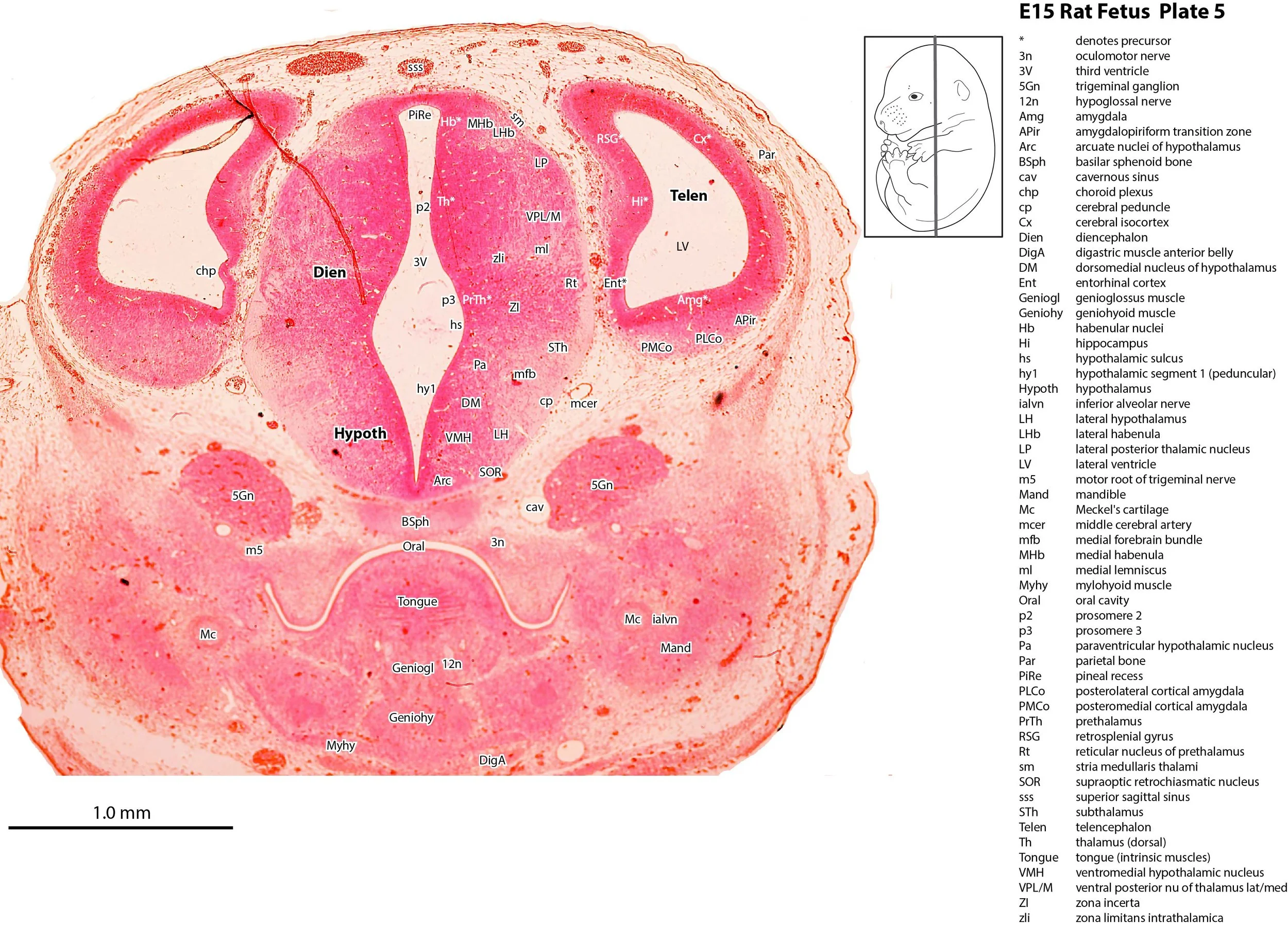
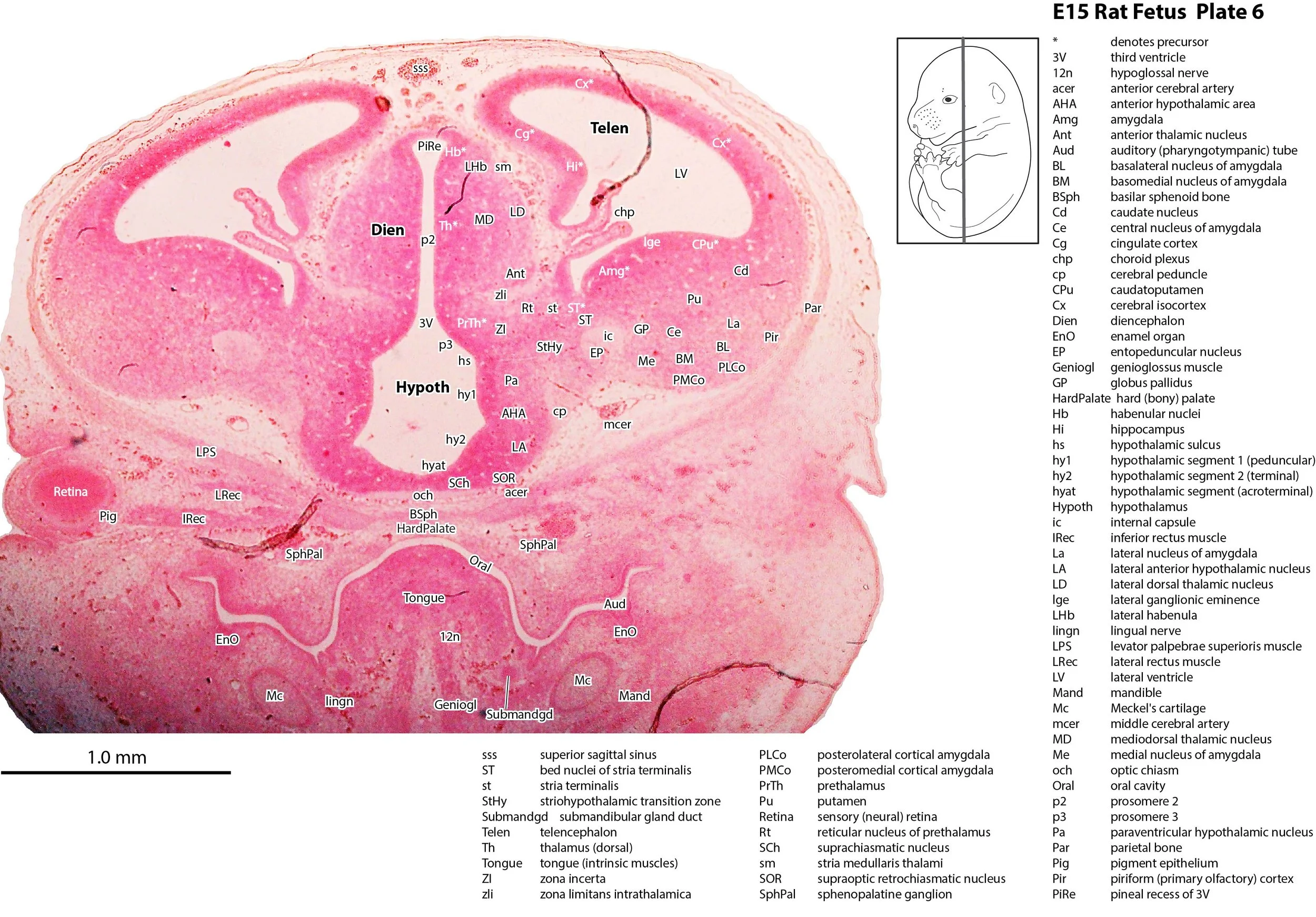
Telencephalon
The cerebral cortex (Cx – Plates 4 to 9) or pallium has begun to show lamination with emergence of a cortical plate in the frontal and parietal regions (CxP in Plates 7 to 9). The hippocampus (Hi – Plate 6) is still very poorly differentiated. Lateral and medial ganglionic eminences (lge, mge in Plates 6 to 8) have appeared, and clumps of striatal and pallidal neurons have been generated (CPu, Cd, Pu, GP, EP, VP in Plates 6 to 8), but detailed topographic separation has yet to be attained. The septal region (Spt in Plate 6) is beginning to show separation into medial and lateral septal nuclei (MS, LS in Plates 7 to 9).
Diencephalon
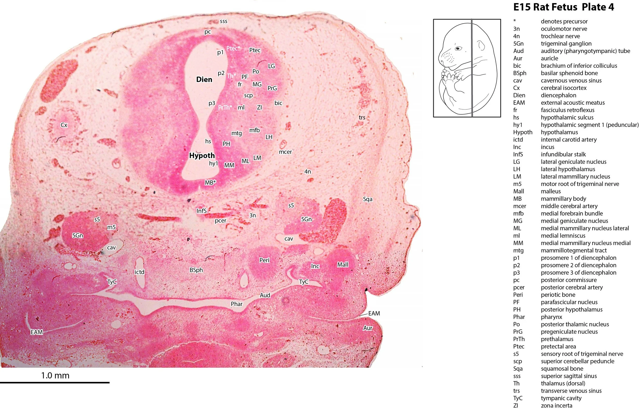
Prosomeres 1 to 3 (Puelles et al. 2012b) are still visible in the diencephalon (see Plates 4 to 6) and neurons are spreading laterally from the diencephalic neuroepithelium into the future pretectum, thalamus and prethalamus (Plates 4 to 6).
Hypothalamus
Developmental subdivisions of the hypothalamus (peduncular, tuberal/terminal and acroterminal hypothalamus; hy1, hy2, hyat, respectively) described by Puelles and co-workers (Puelles et al. 2012a) have been identified here (Plates 4 to 6).
Cerebellum
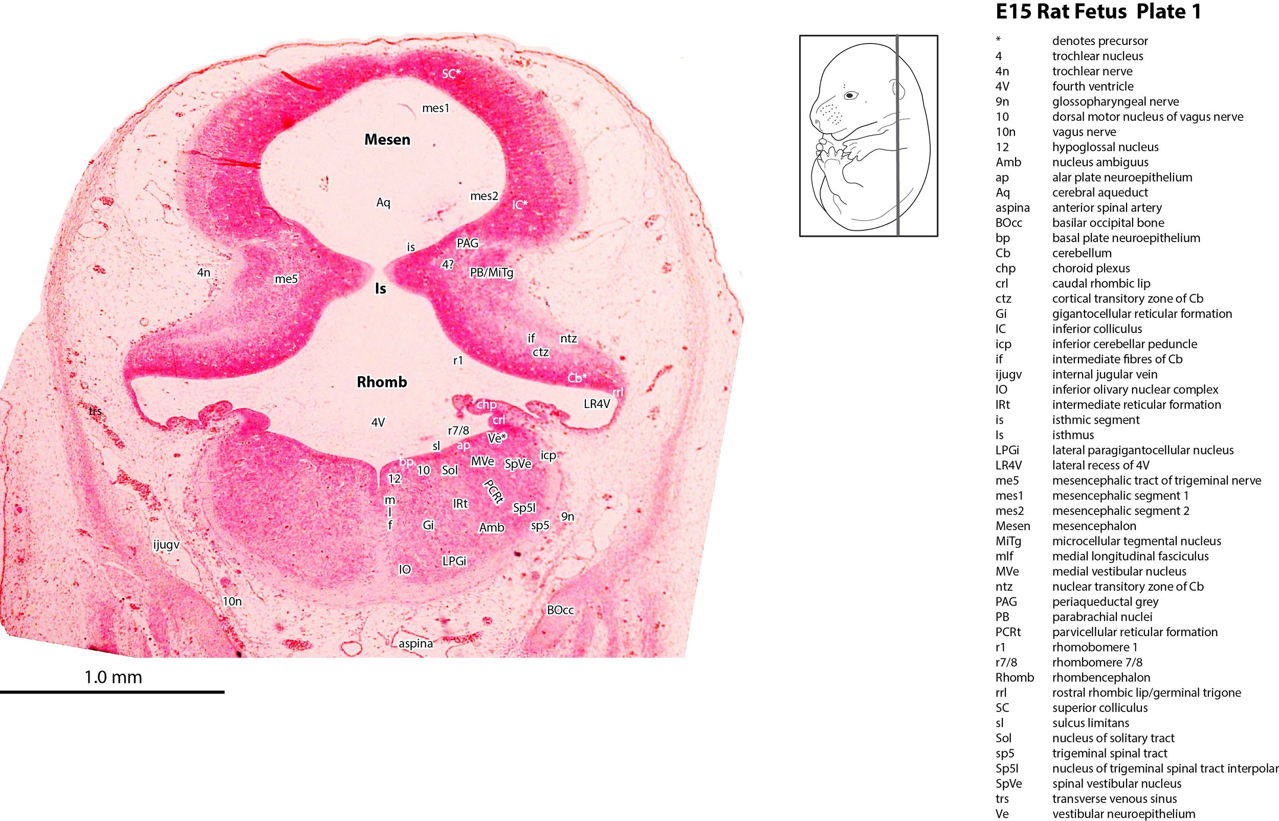
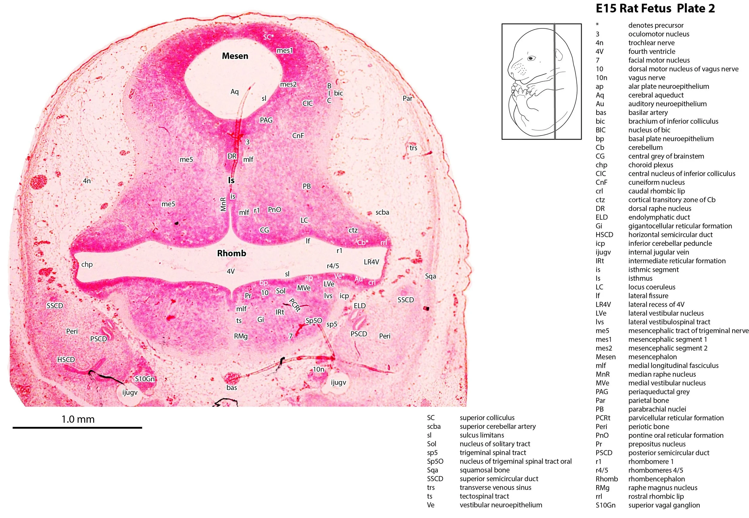
The rostral rhombic lip (rrl in Plates 1 and 2) forming the roof of the fourth ventricle has expanded to form a cerebellar shelf. Cerebellar macroneuron neurogenesis has begun and cortical and nuclear transitory zones are visible (ctz, ntz in Plates 1 and 2) separated by intermediate fibres (if in Plate 1).
Brainstem
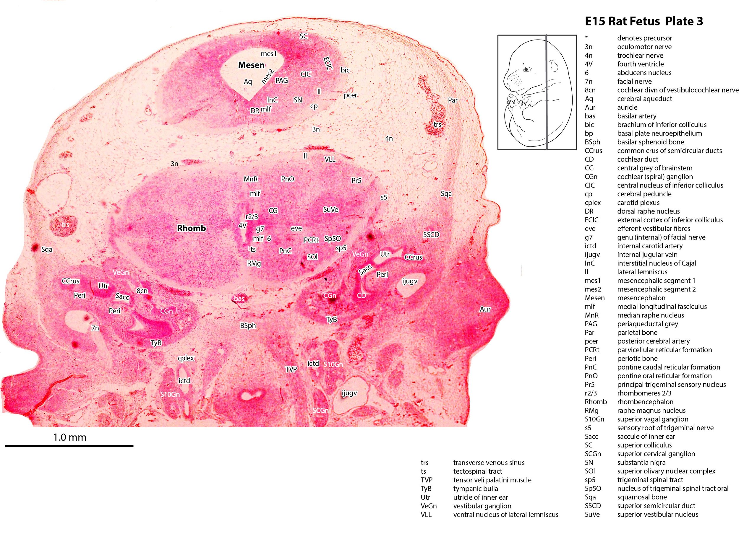
Alar and basal plates are still visible separated by the sulcus limitans. Major motor and sensory nuclei have begun to settle adjacent to the pial membrane, but separation of individual nuclei is difficult.
References
Puelles L, Martinez-de-la-Torre M, Bardet S and Rubenstein JLR (2012a) Hypothalamus. In The Mouse Nervous System(Eds C Watson, G Paxinos and L Puelles) pp. 221–312. Elsevier, San Diego.
Puelles L, Martinez-de-la-Torre M, Ferran J-L and Watson CRR (2012b) Diencephalon. In The Mouse Nervous System(Eds C Watson, G Paxinos and L Puelles) pp. 313–336. Elsevier, San Diego.