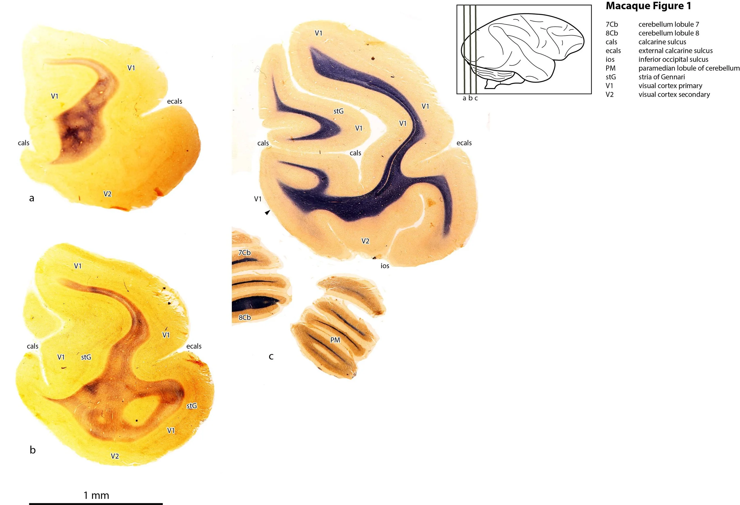
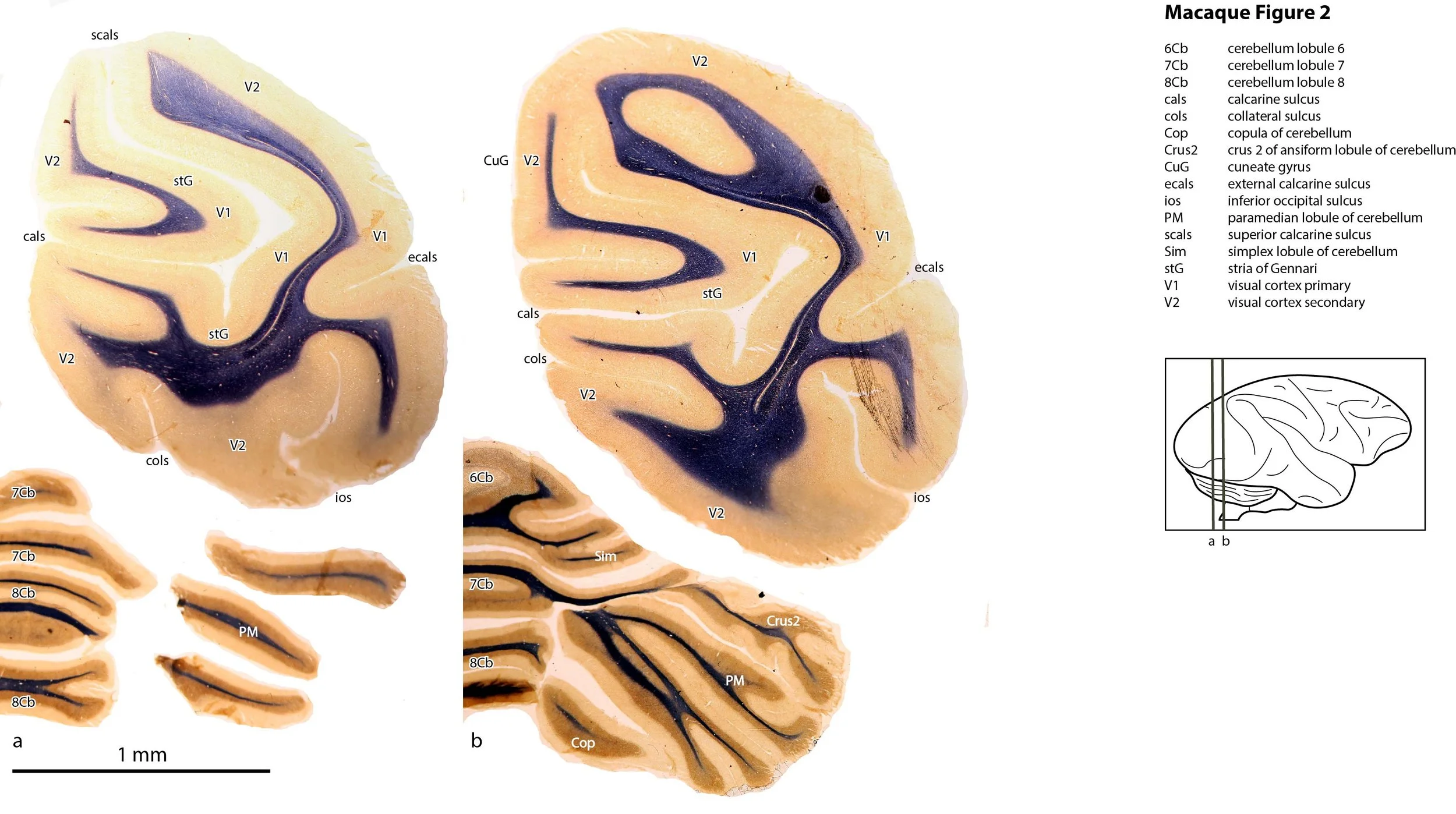
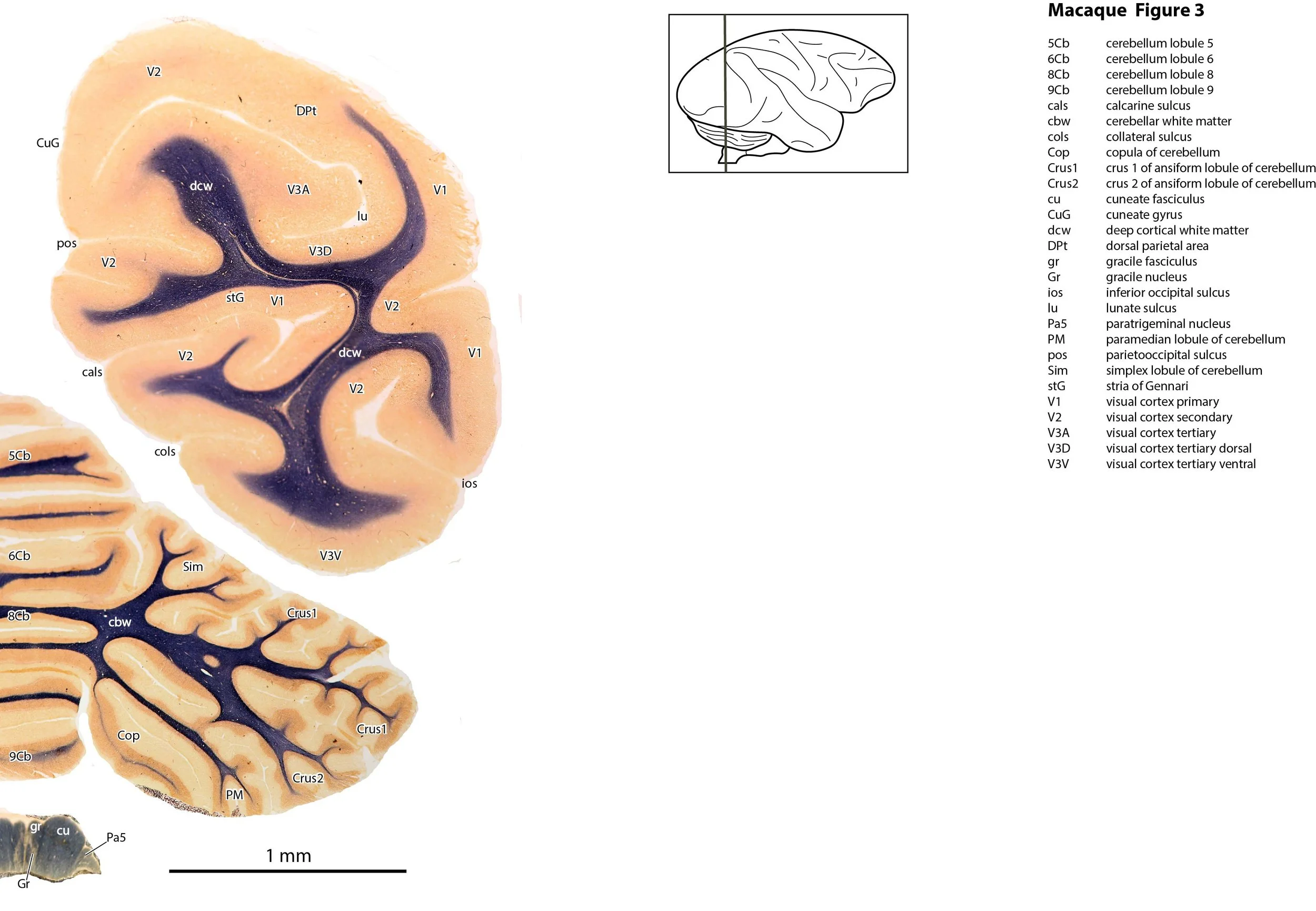
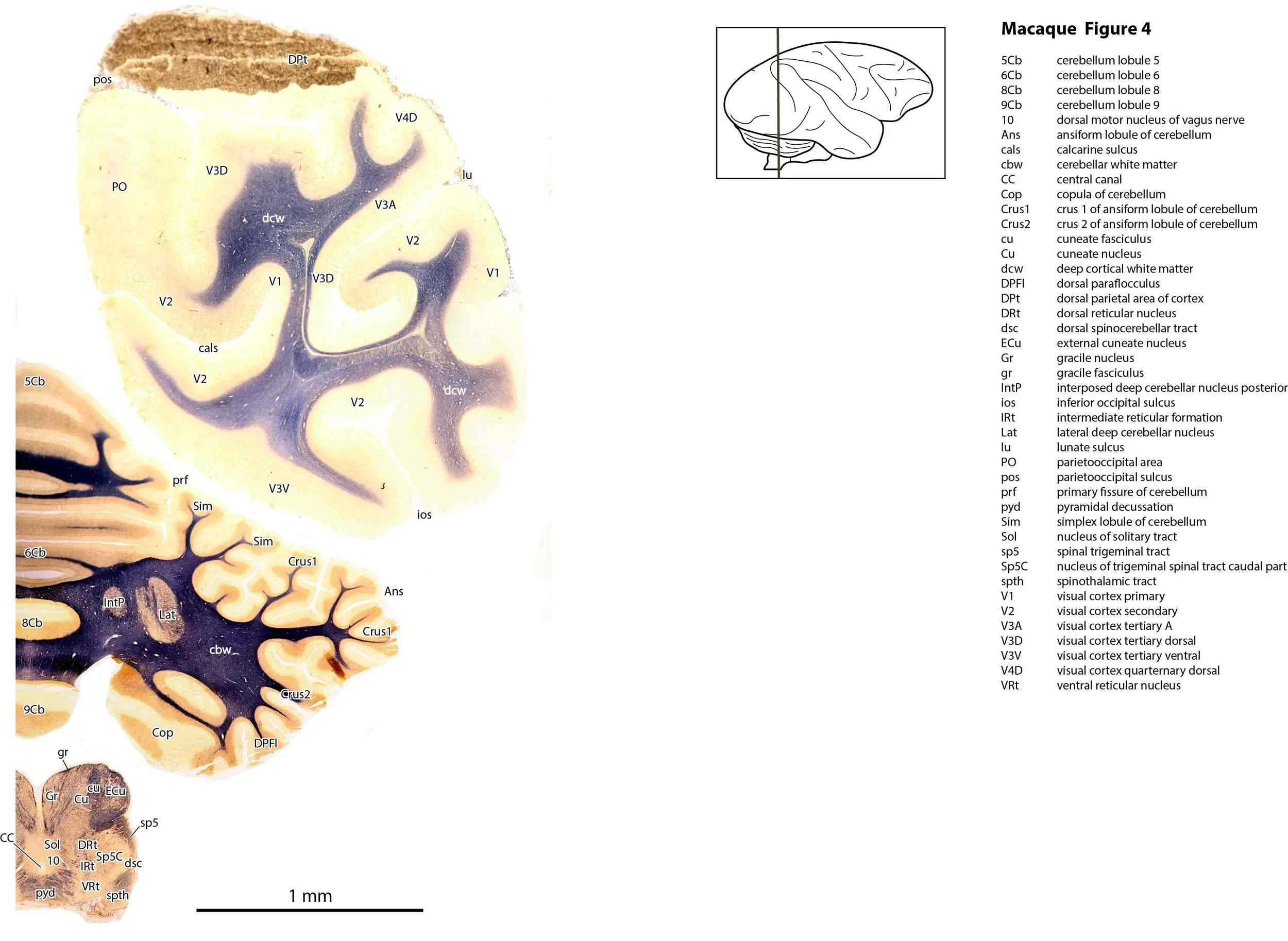
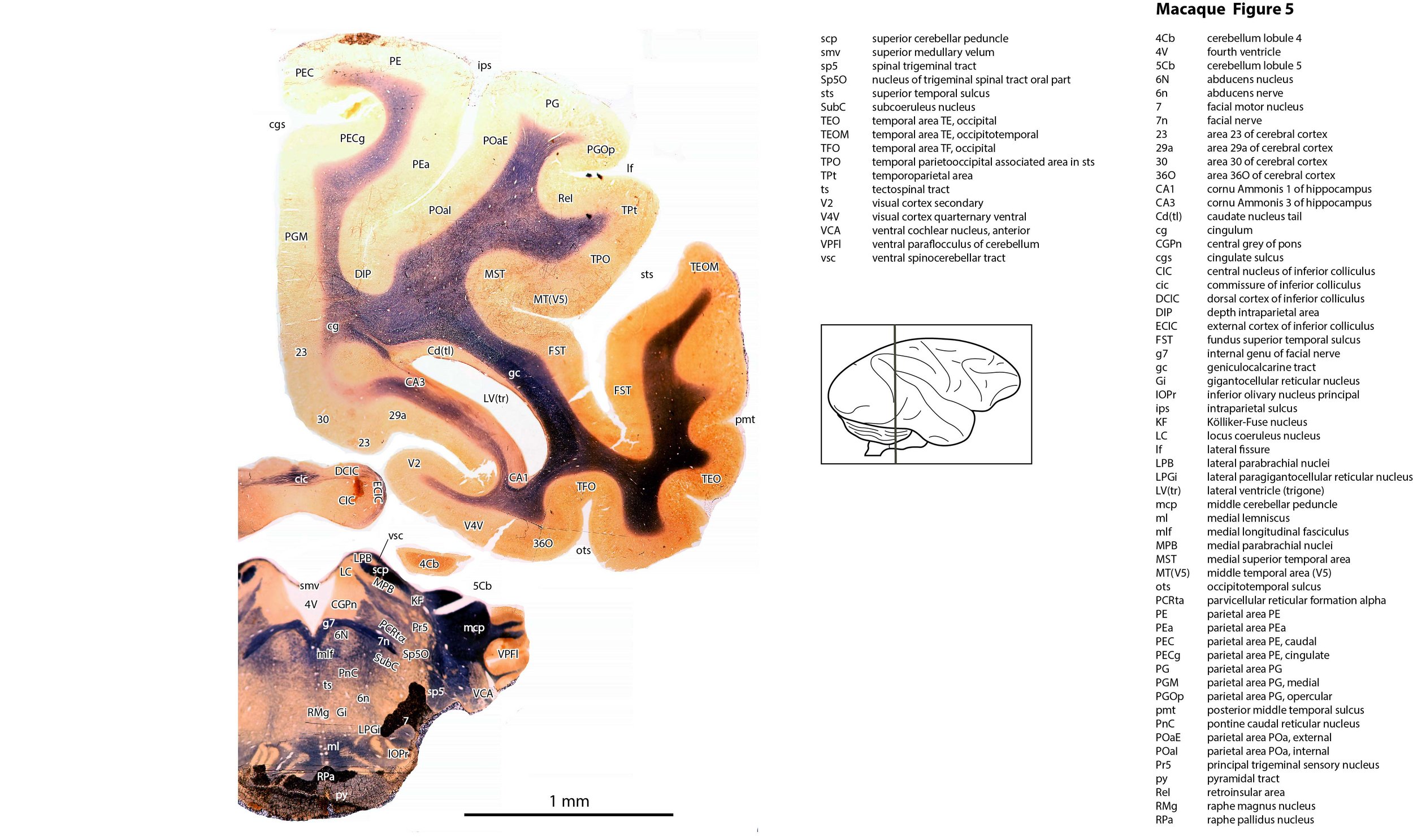
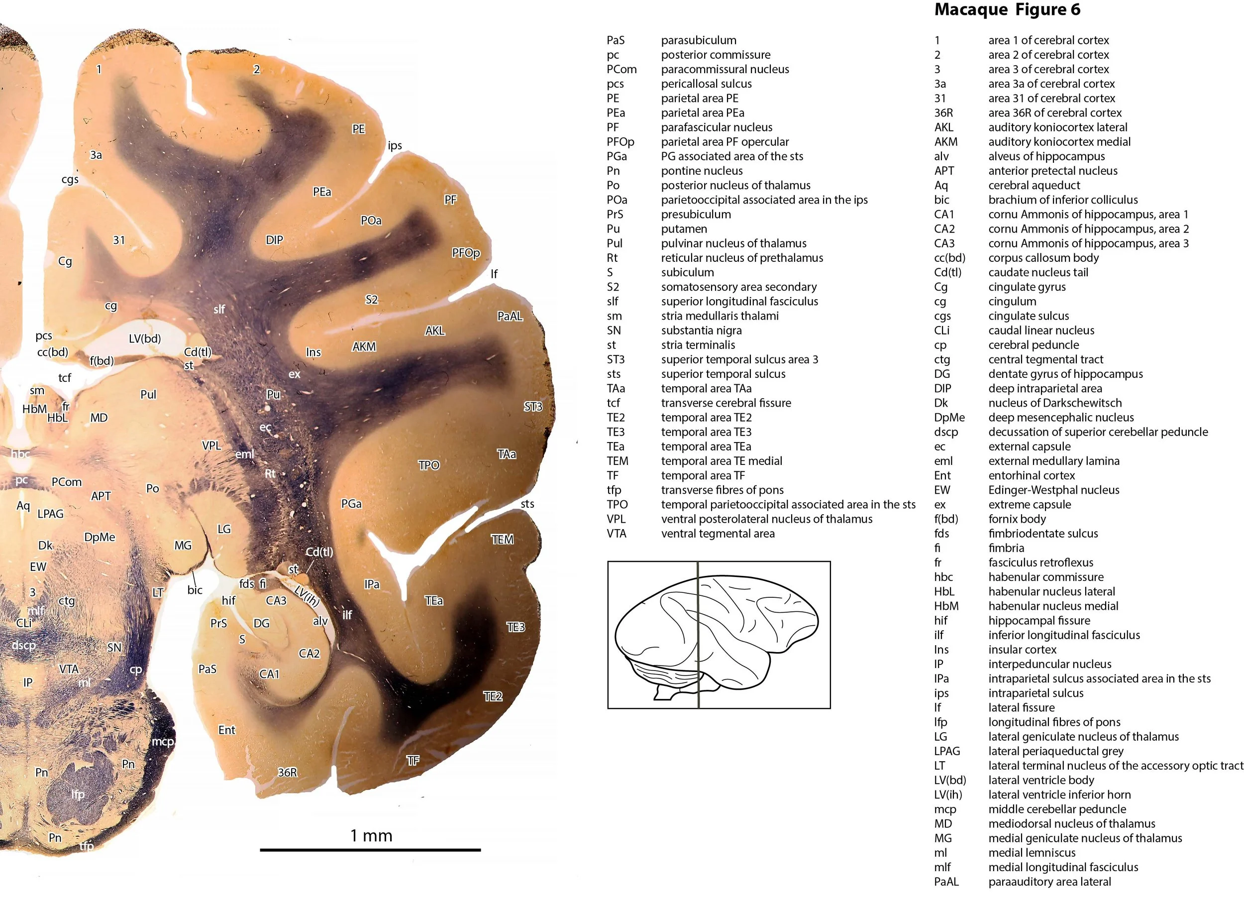
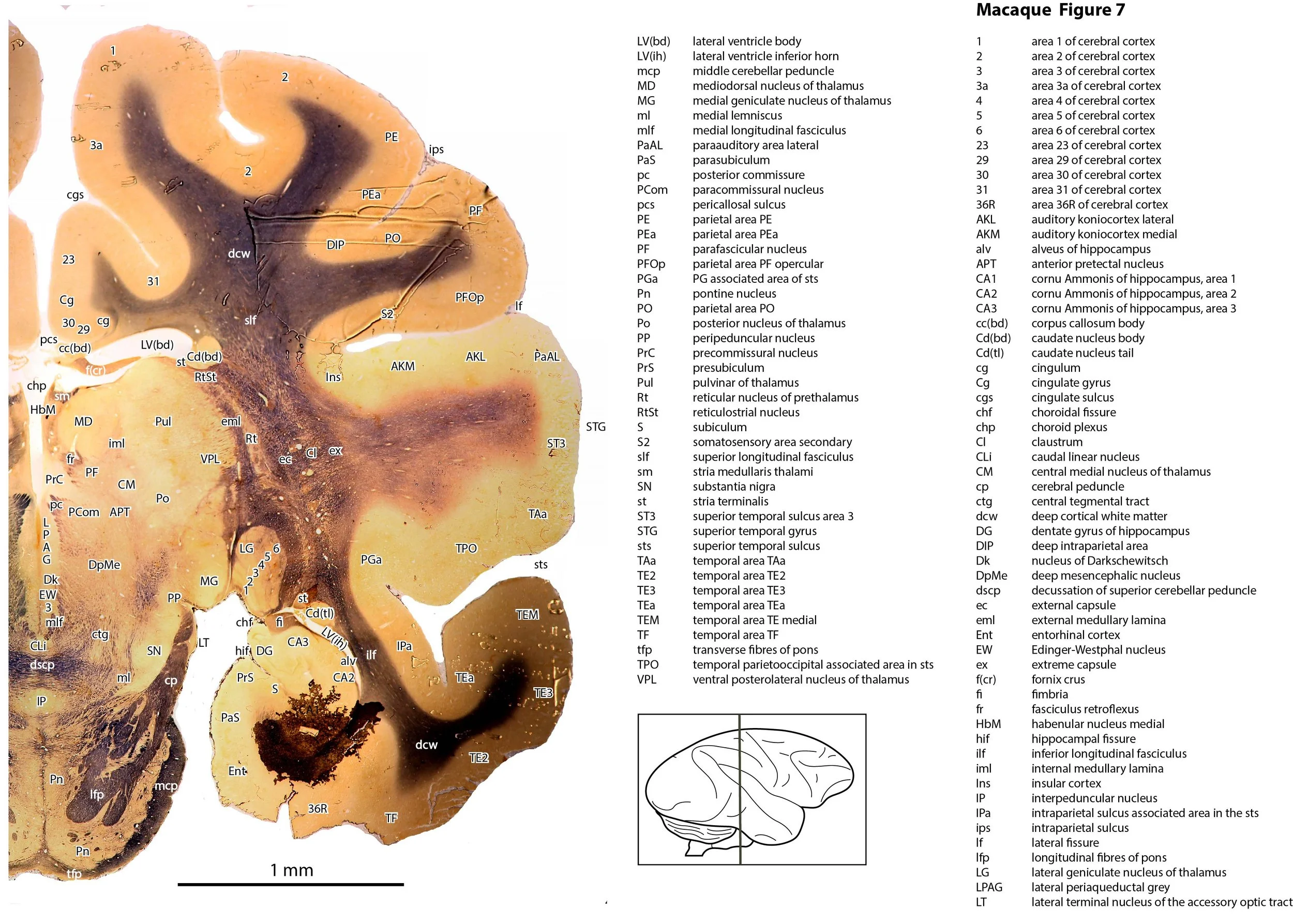
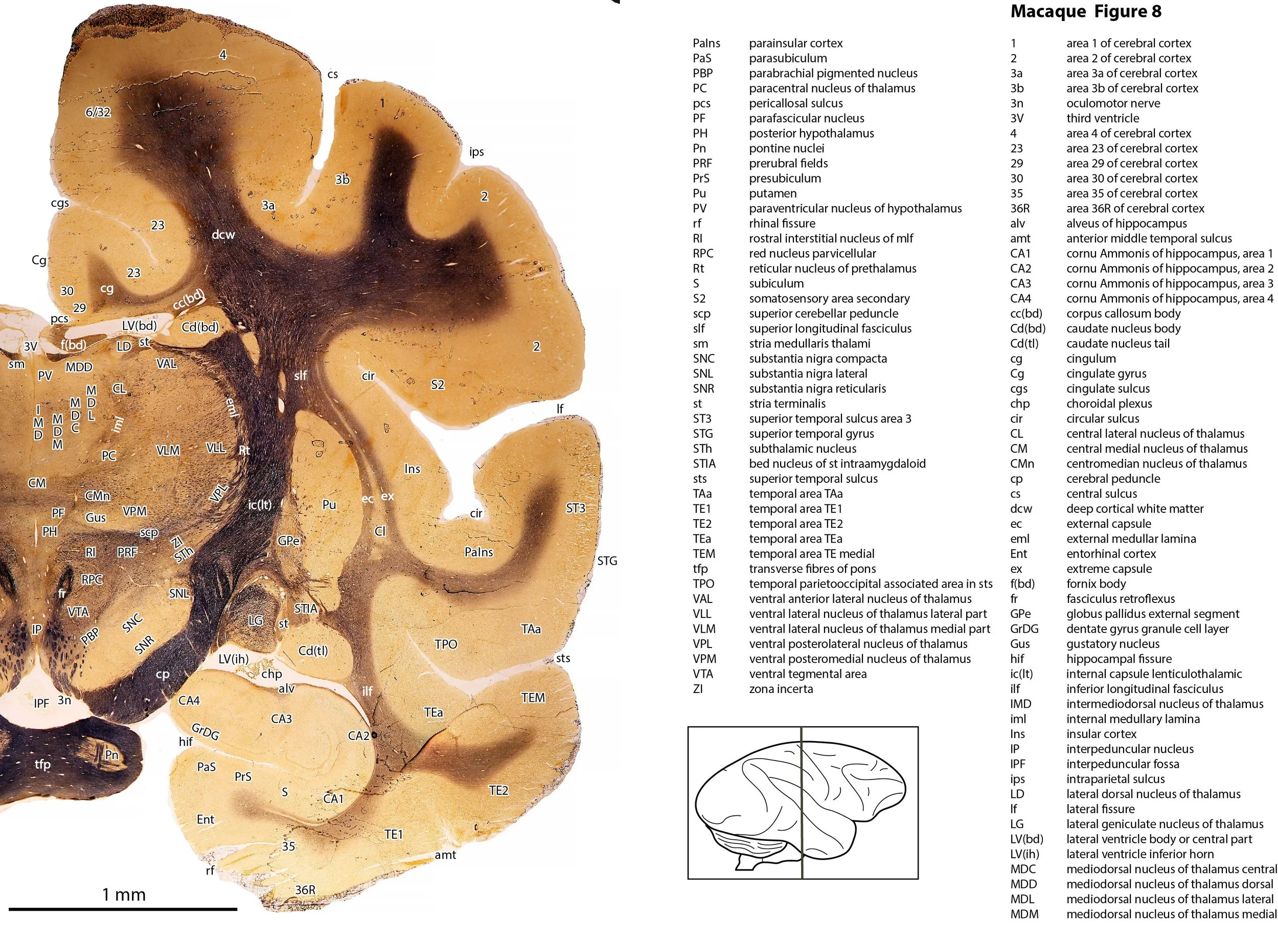
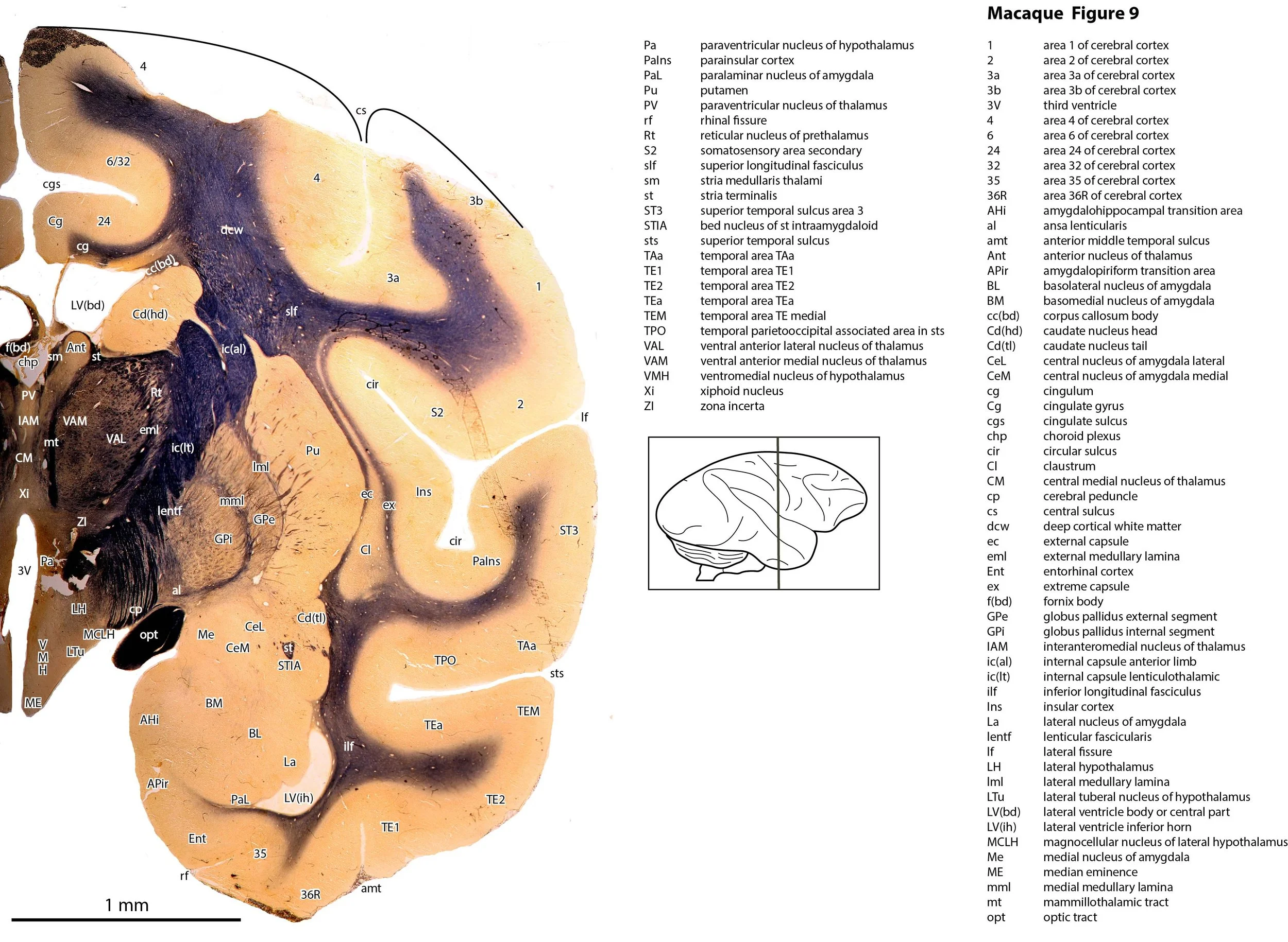
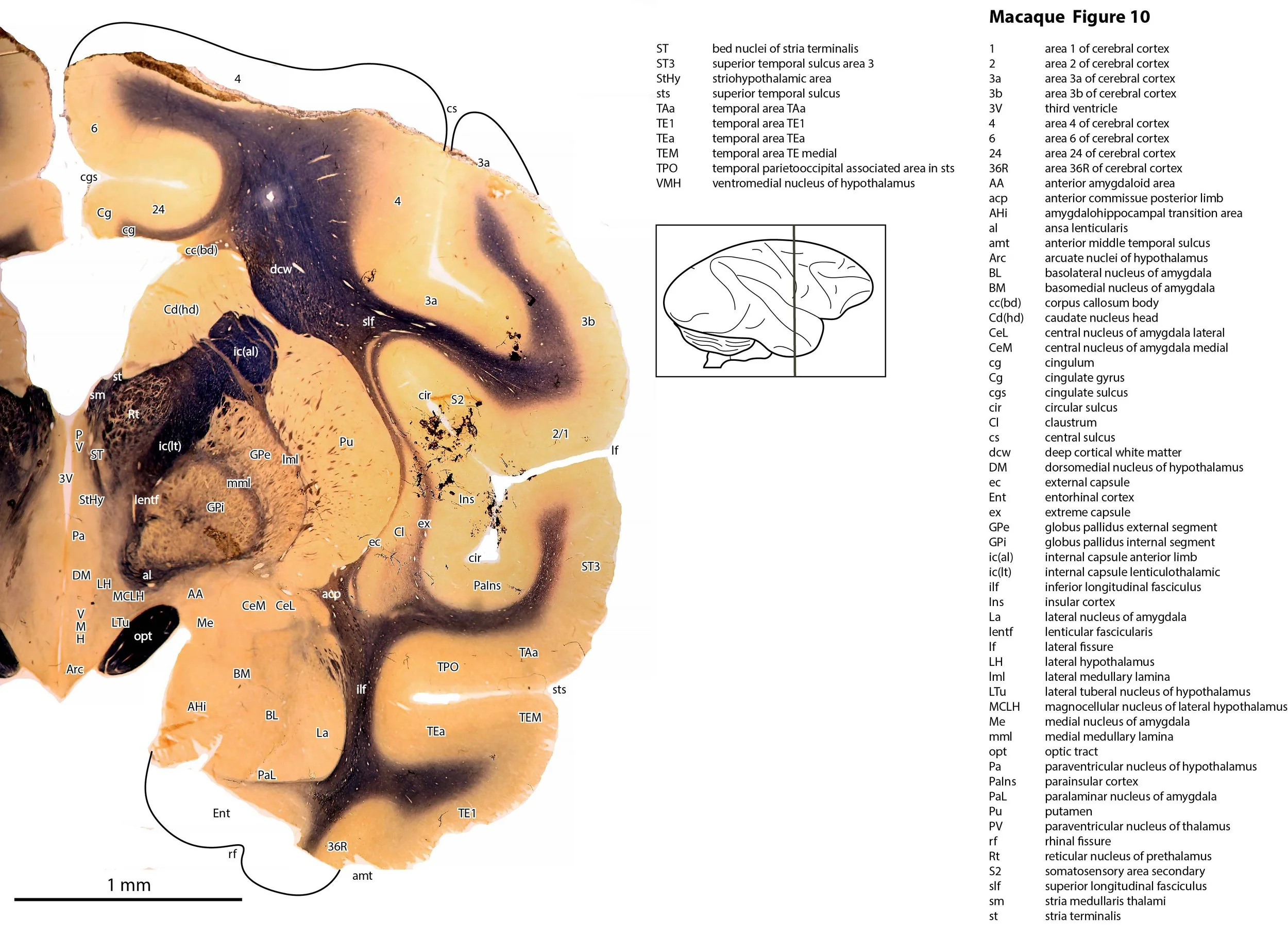
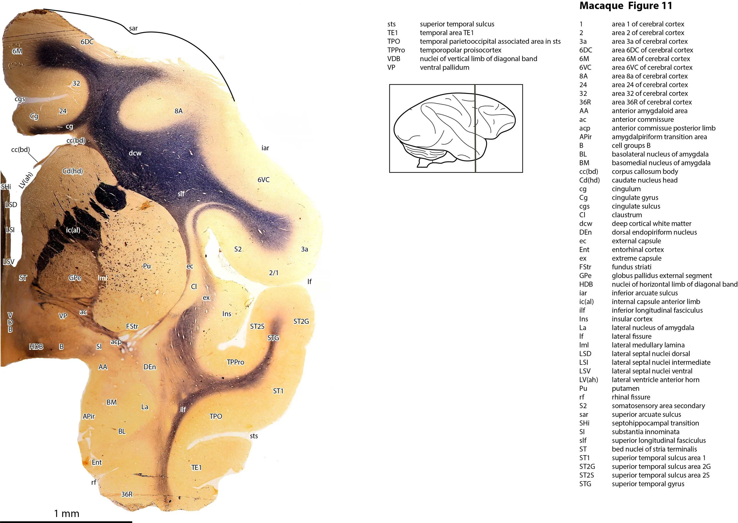
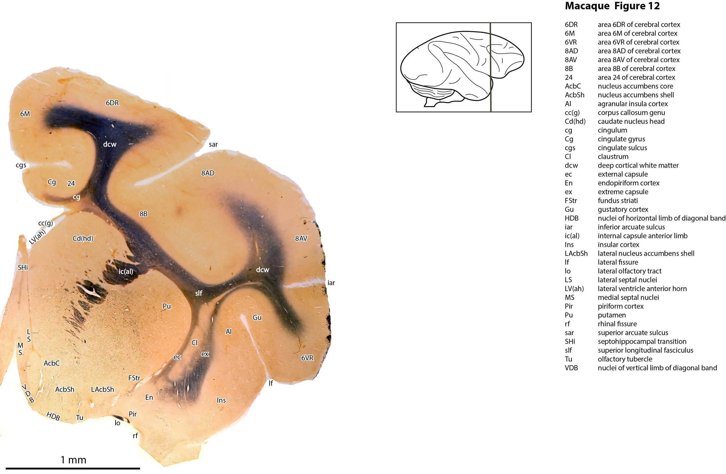
Atlas of the Brain of a Macaque
This specimen was kindly donated by Dr Lauren Marotte from the collection of the late Professor Richard Mark of the Research School of Biological Sciences, Australian National University. The animal (Topsy) had been used in a study of bimanual co-ordination in monkeys after corpus callosum and anterior commissure transection (Mark & Sperry, 1968). For this reason, the corpus callosum is damaged in the sections, but still visible as a stub. Nevertheless, all other parts of the brain are intact and can be used to study normal primate neuroanatomy.
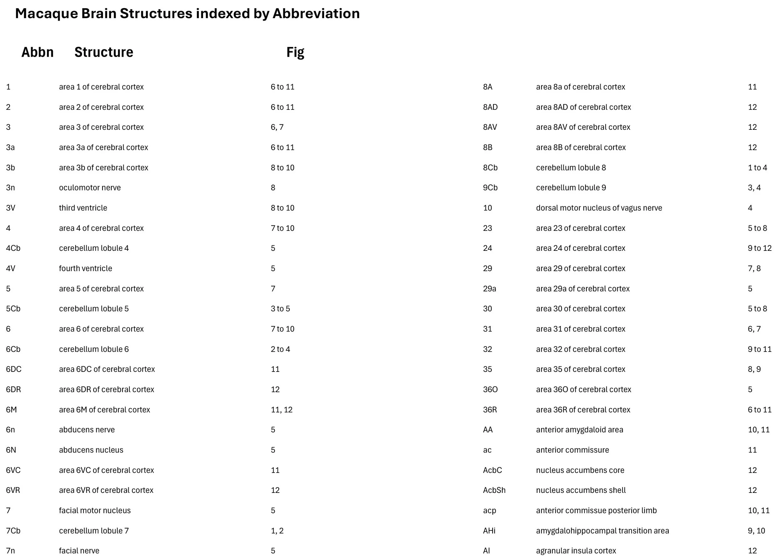
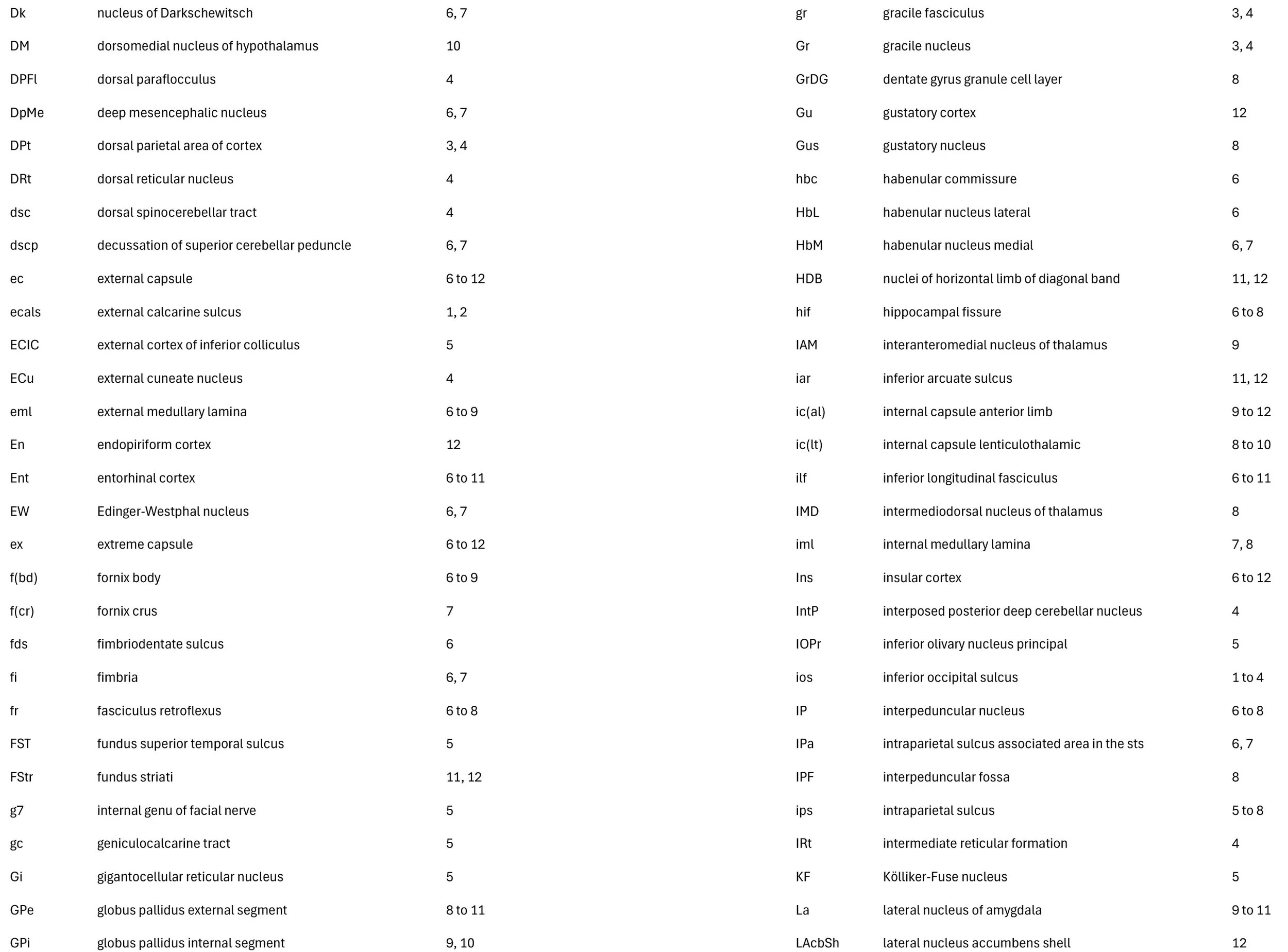
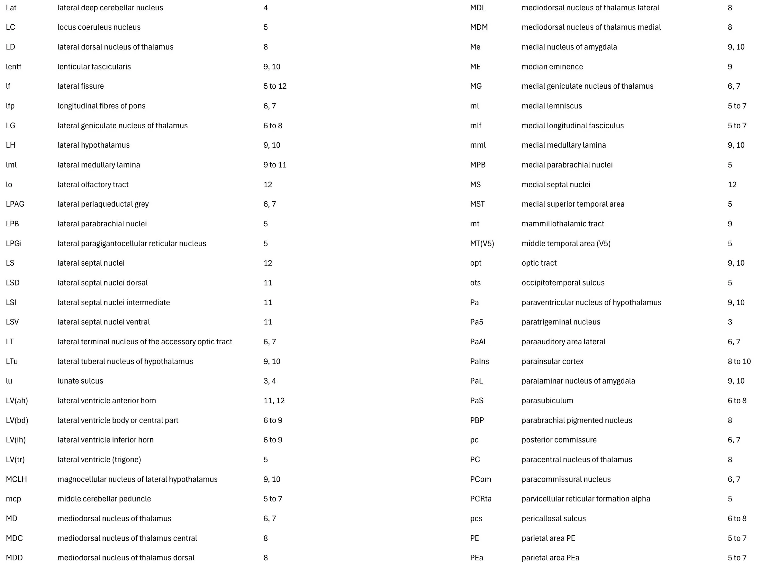
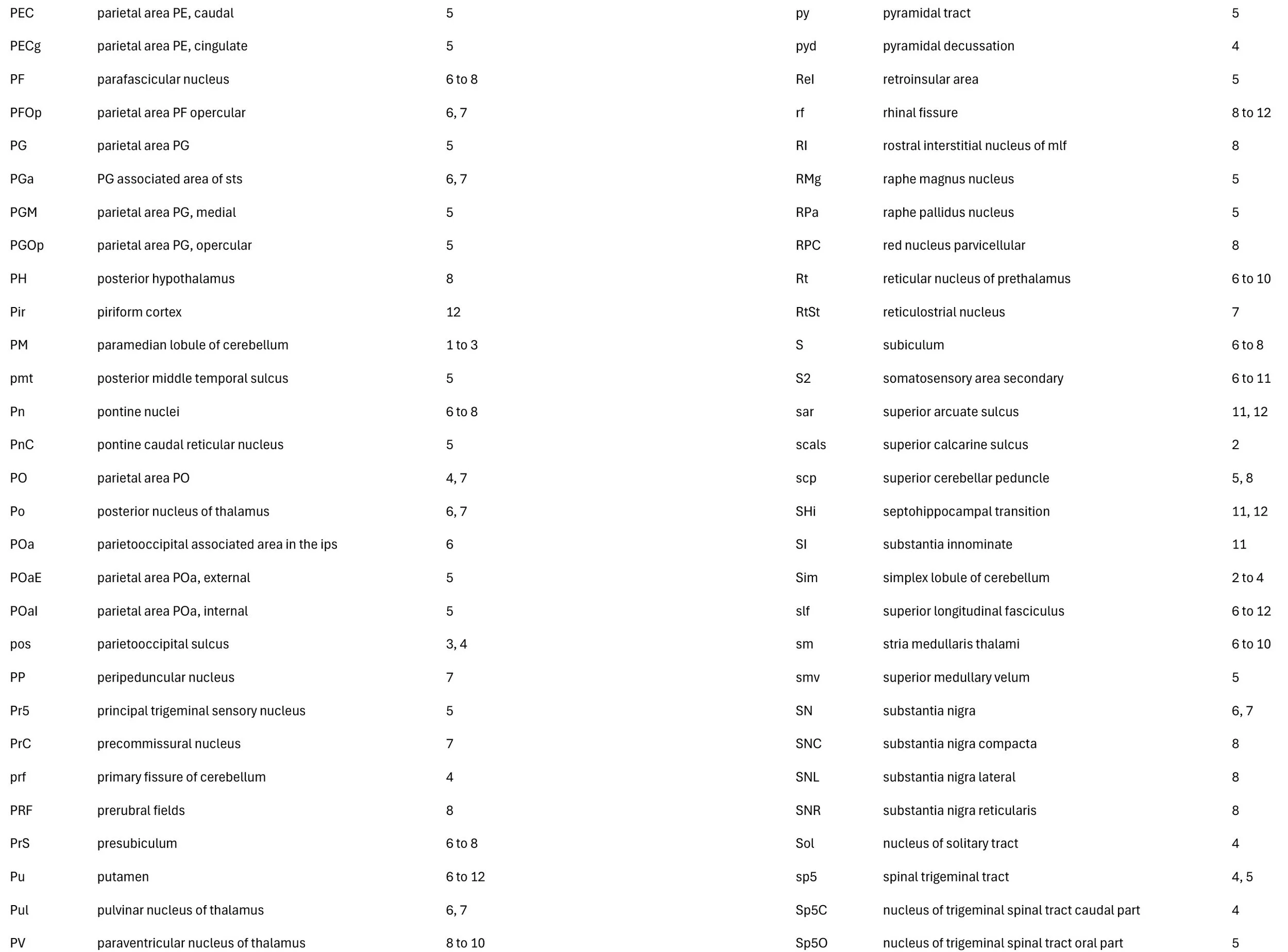
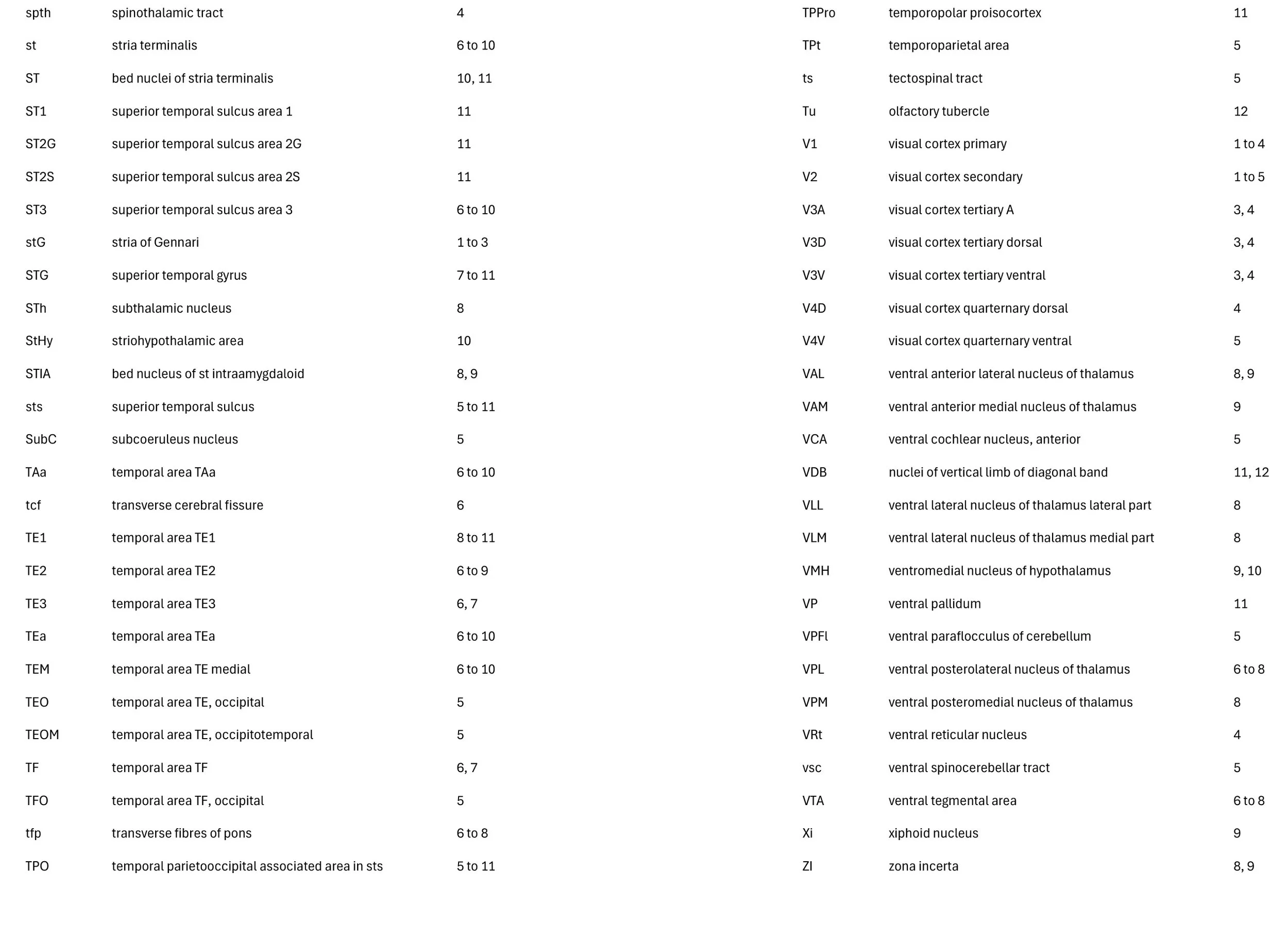
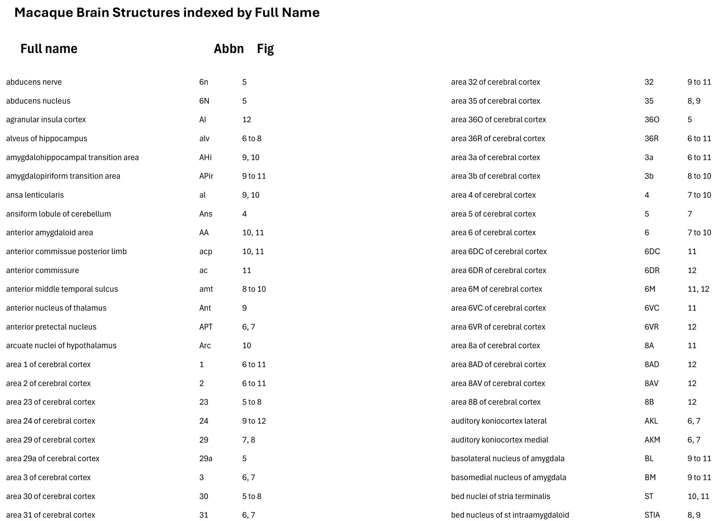
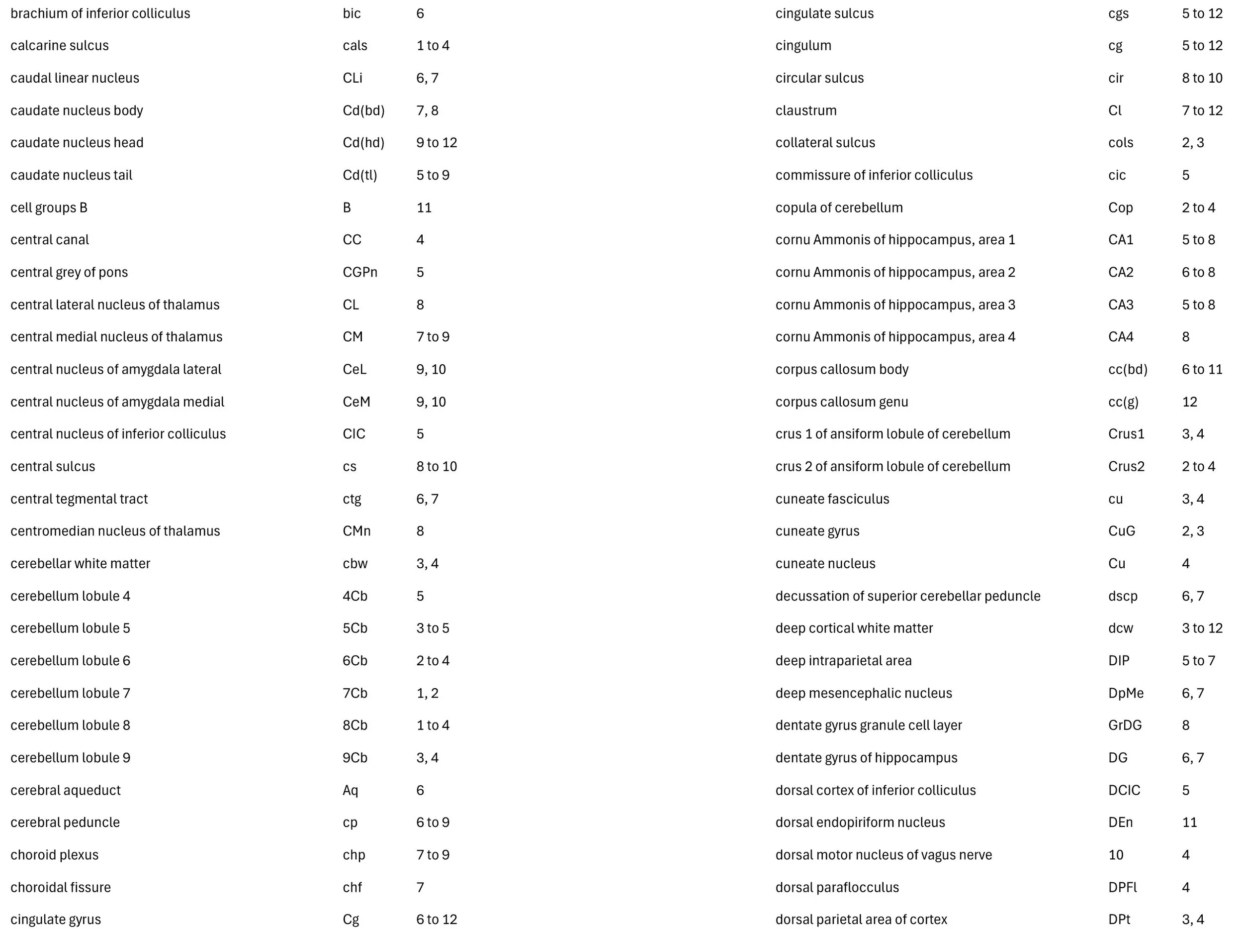
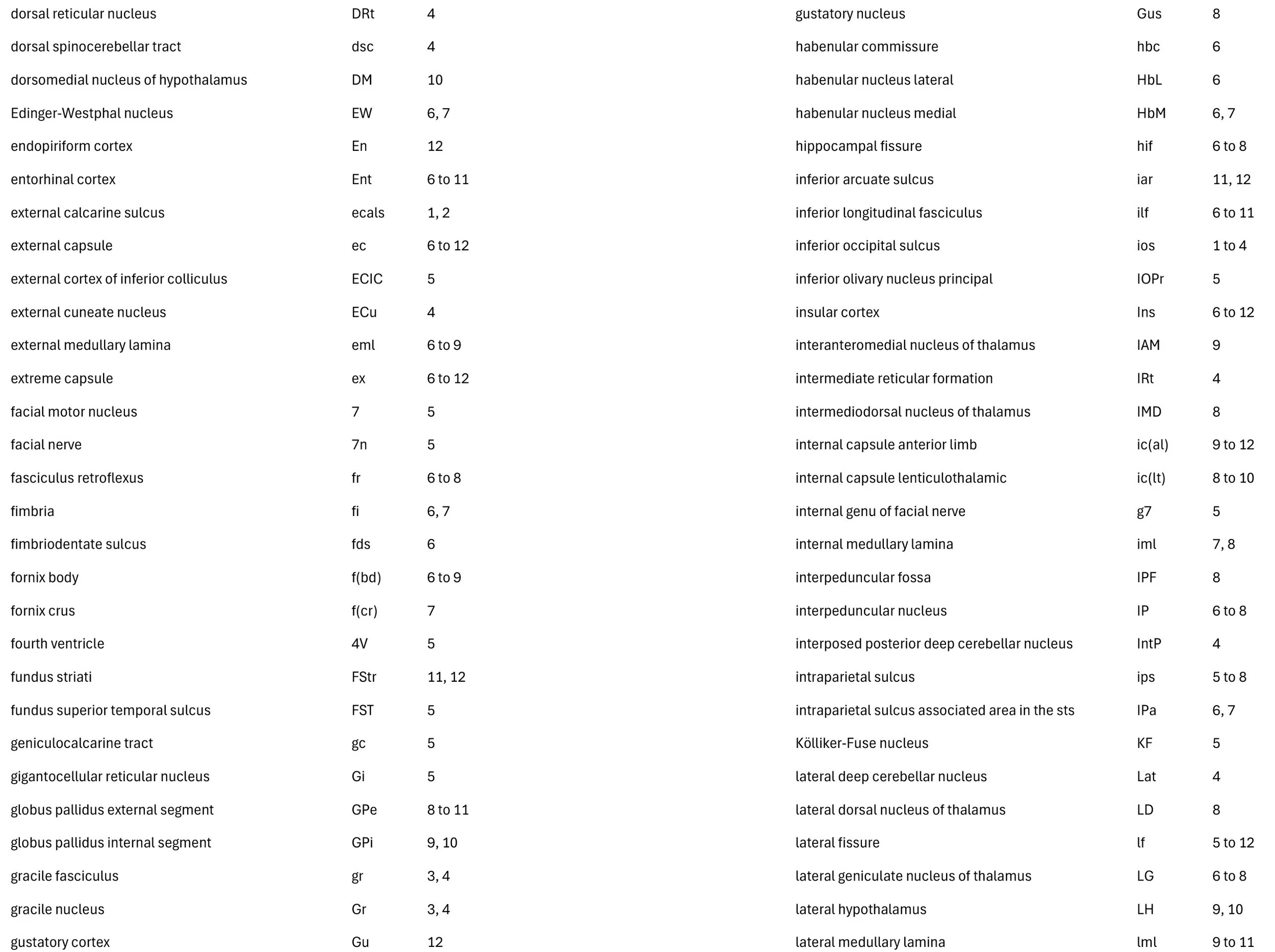
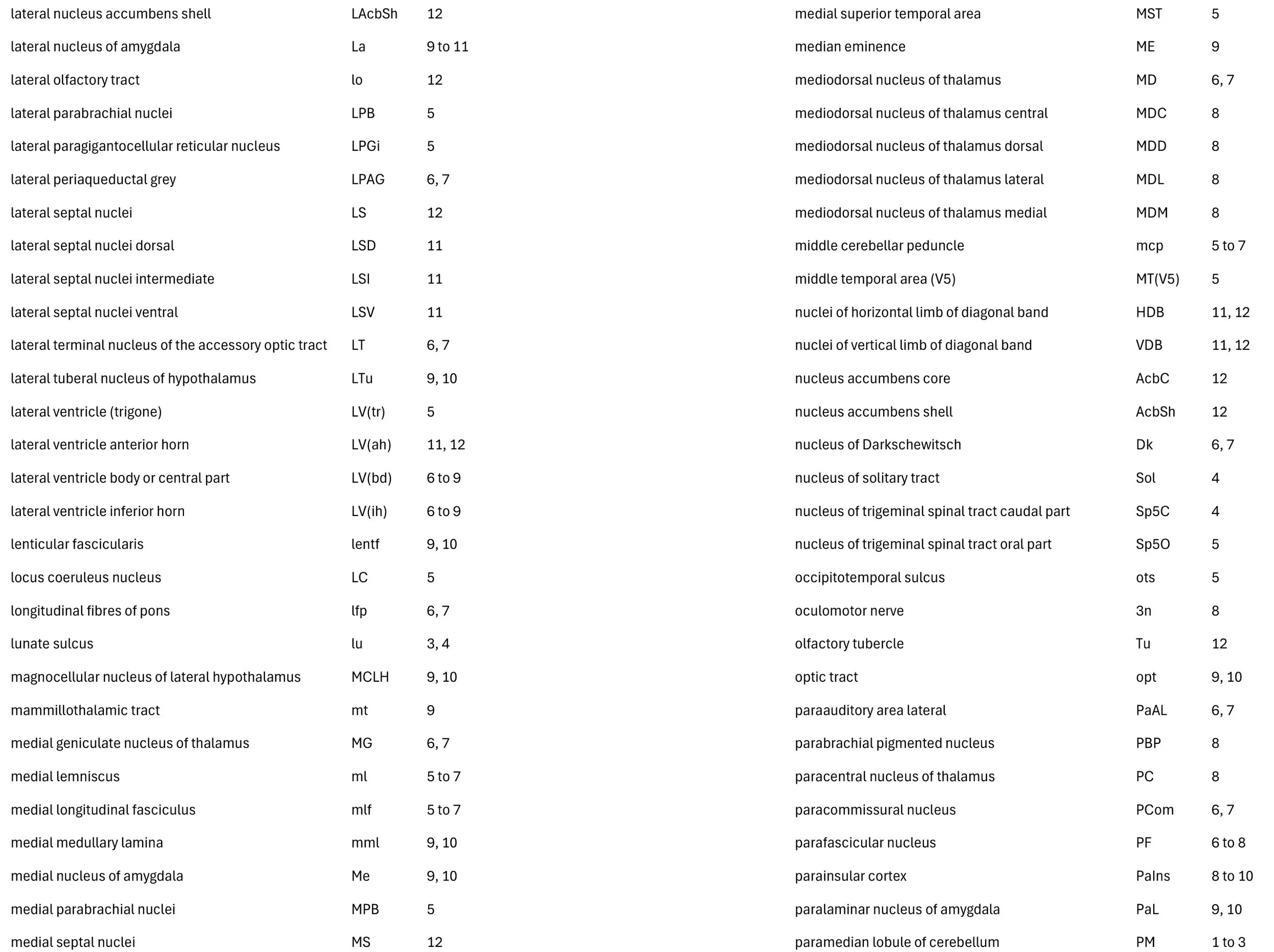
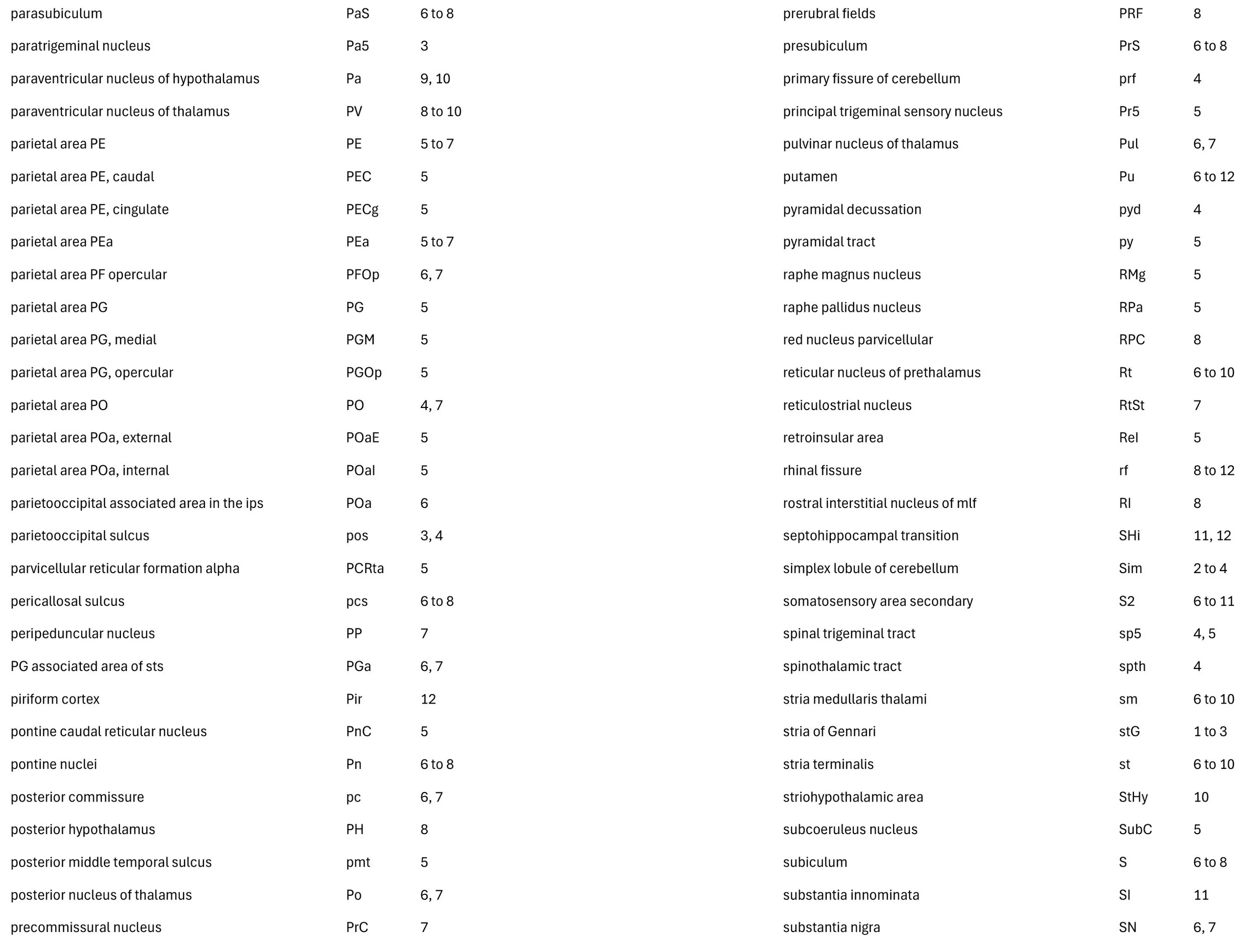
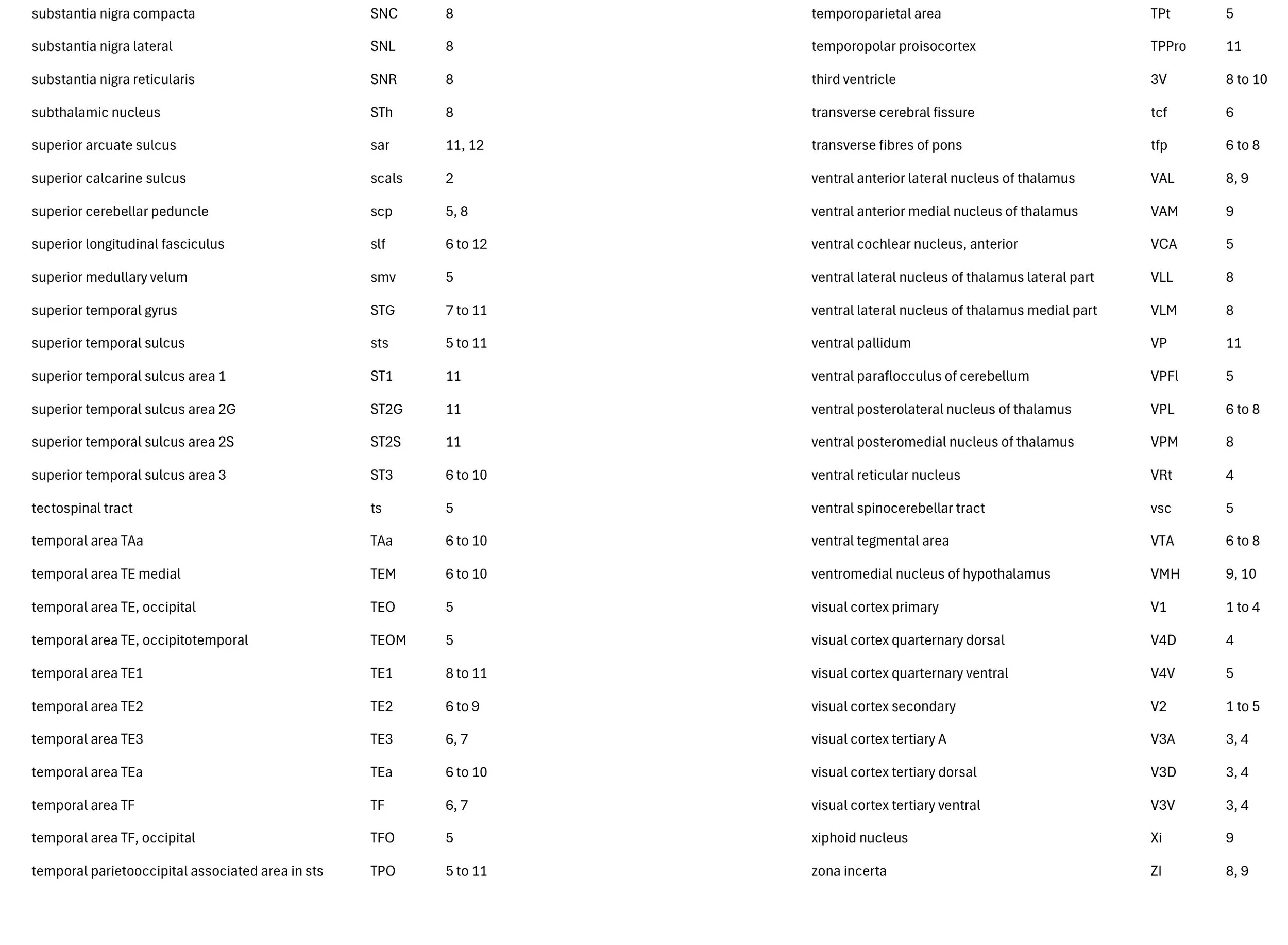
The right side of the brain is shown in all images. The specimen sections had been treated with a variant of the Weil myelin stain that shows myelinated fibres as dark and nuclei as pale. Boundaries between cortical areas are not distinguishable, so notional positions of cortical regions based on Paxinos et al. (2000) have been made. Nomenclature for cortical gyri and sulci also follow Paxinos et al. (2000).
References
Mark RF, Sperry RW (1968) Bimanual coordination in monkeys. Expl Neurol 21: 92-104.
Paxinos G, Huang X-F, Toga AW (2000) The Rhesus Monkey Brain in Stereotaxic Co-ordinates. Academic.