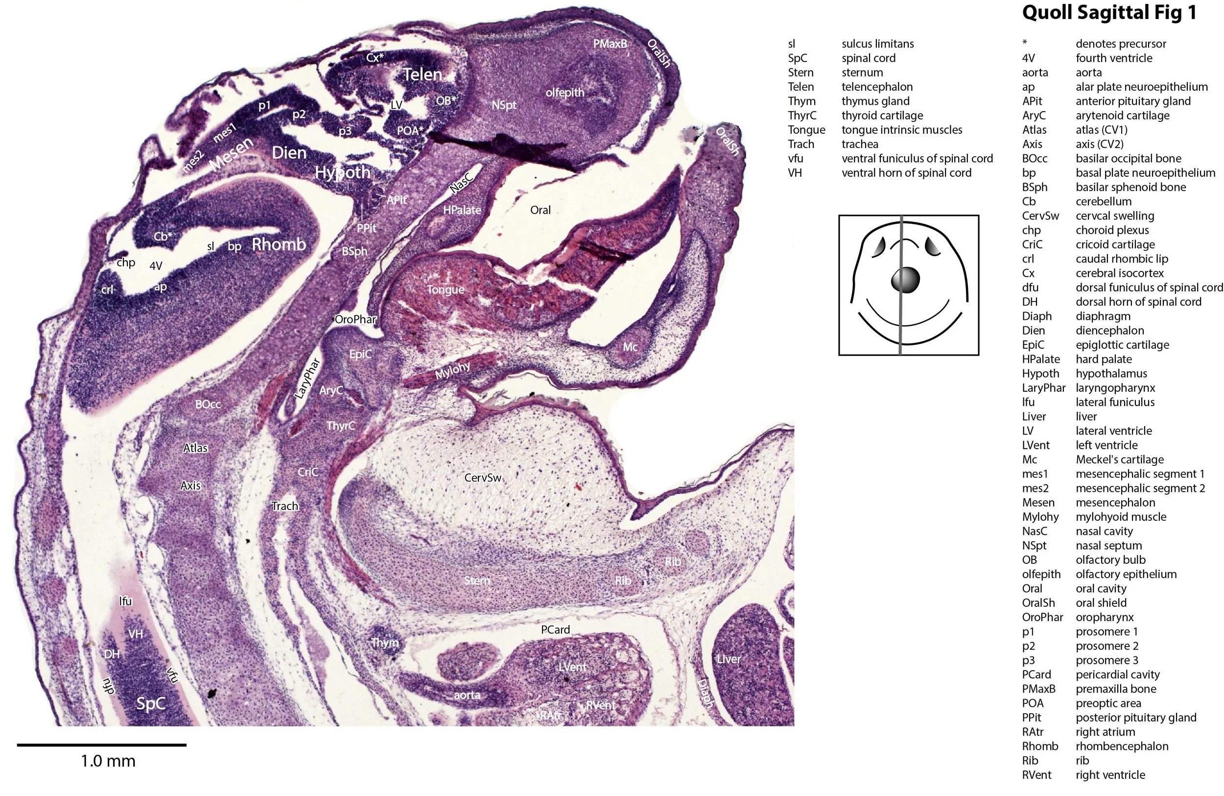
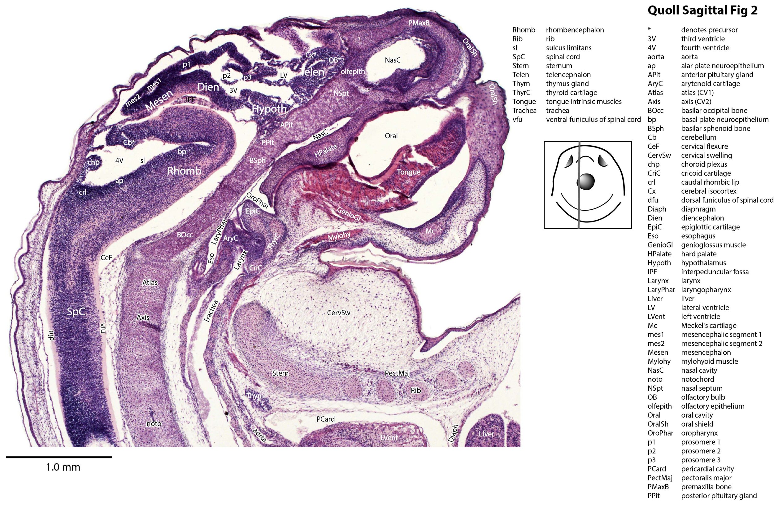
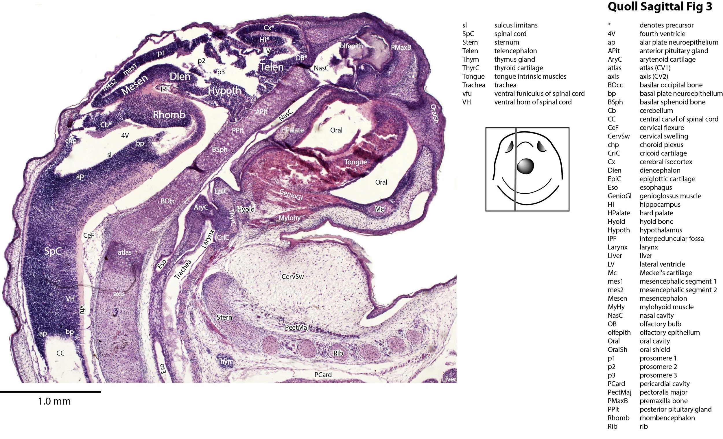
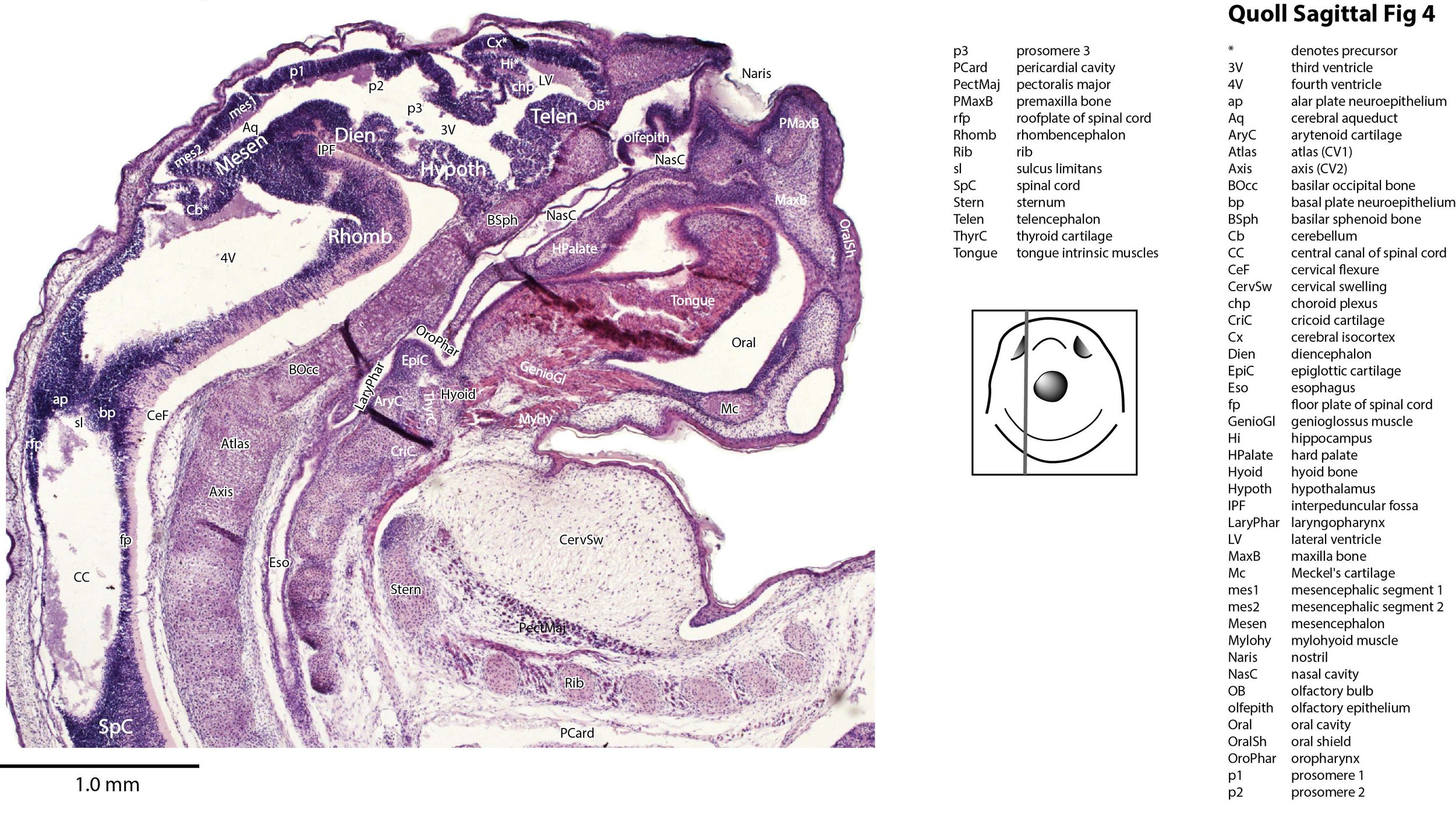
Newborn Eastern Quoll Head and Neck Sagittal Sections (MS148b)
Introduction
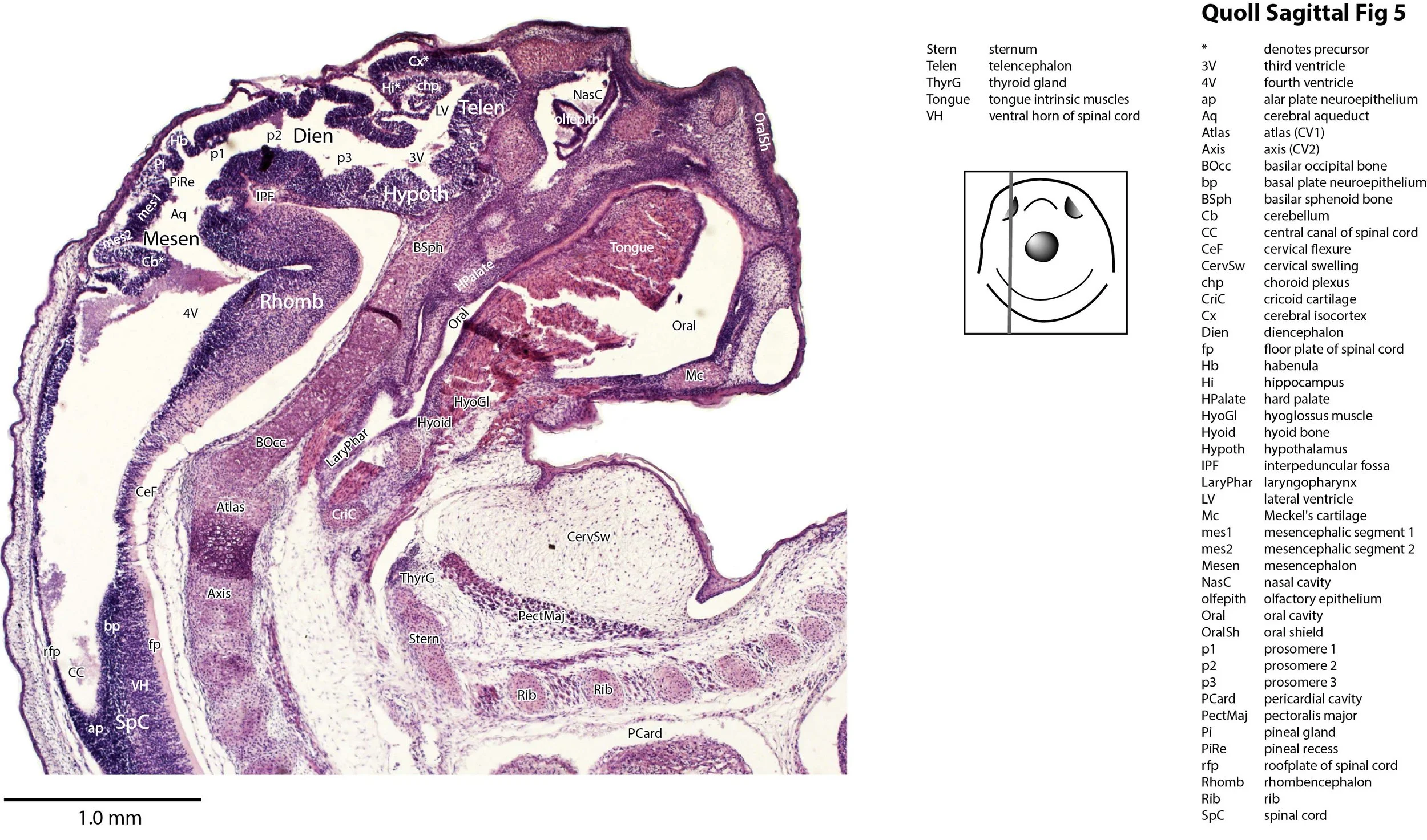
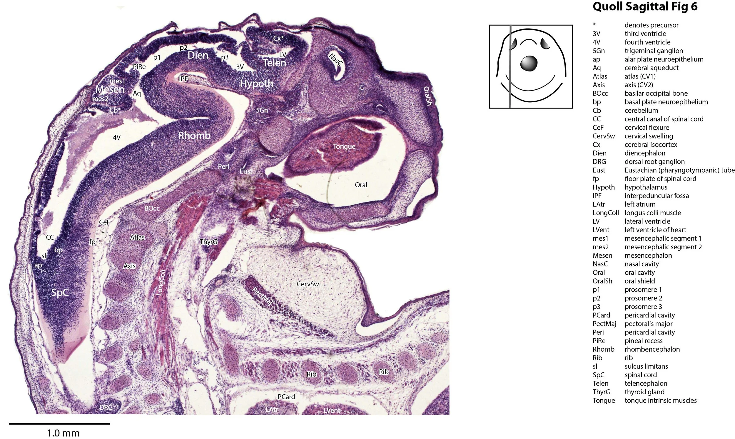
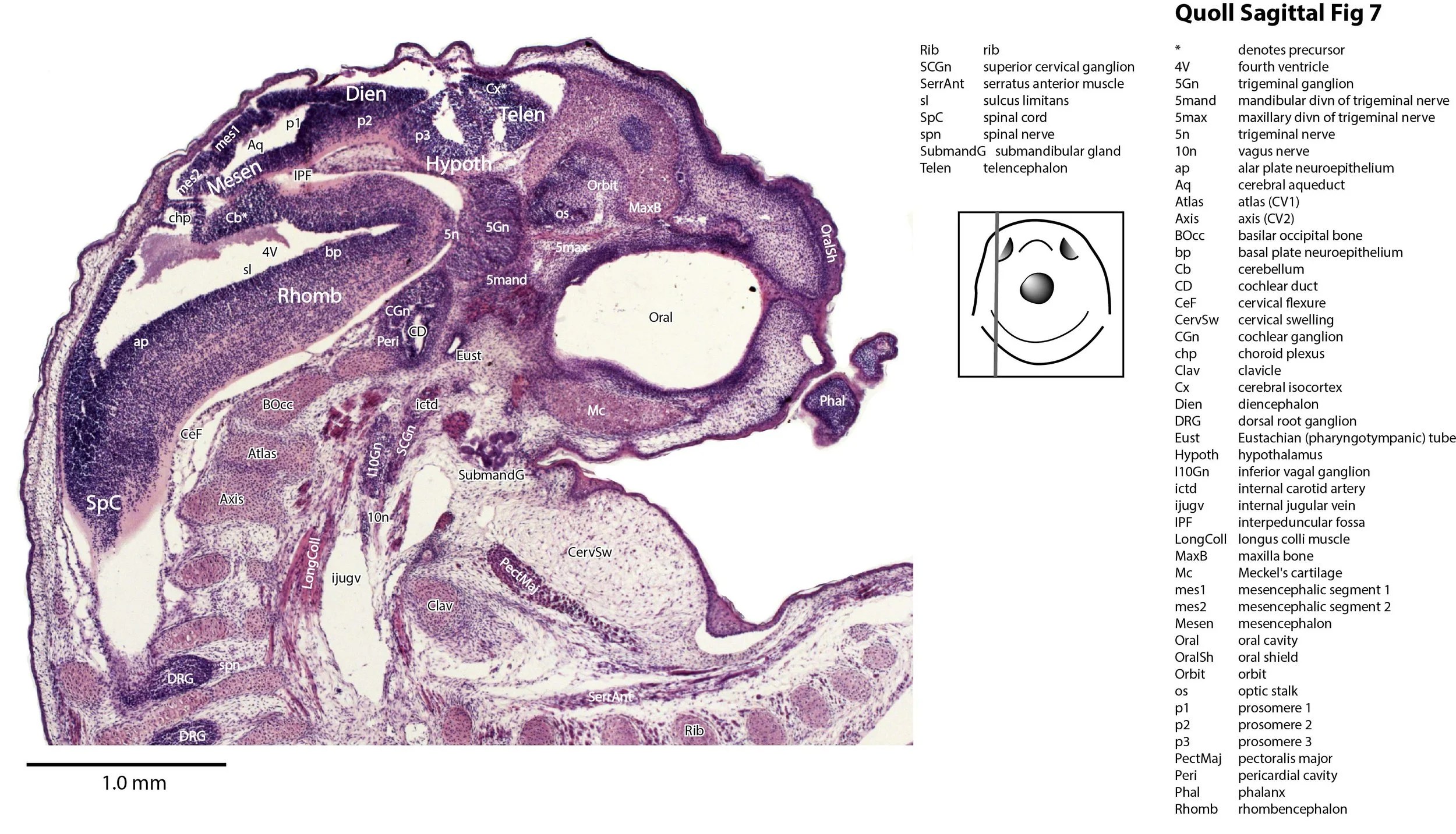
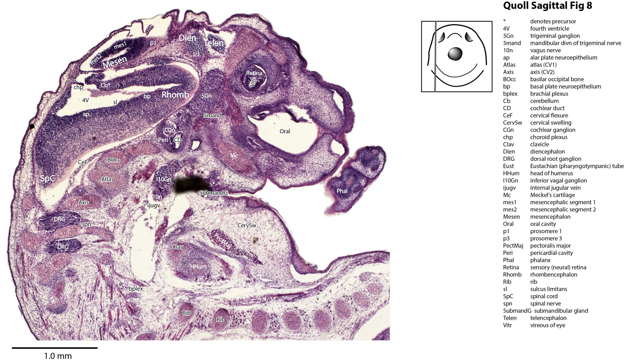
This specimen is a newborn Dasyurus viverrinus pouch young. The specimen is MS148b of the Hill Collection held at the Museum für Naturkunde in Berlin (7.0 mm GL and 4.0 mm HL). Please see the horizontally sectioned dasyurid newborn for details of this species.
Methods
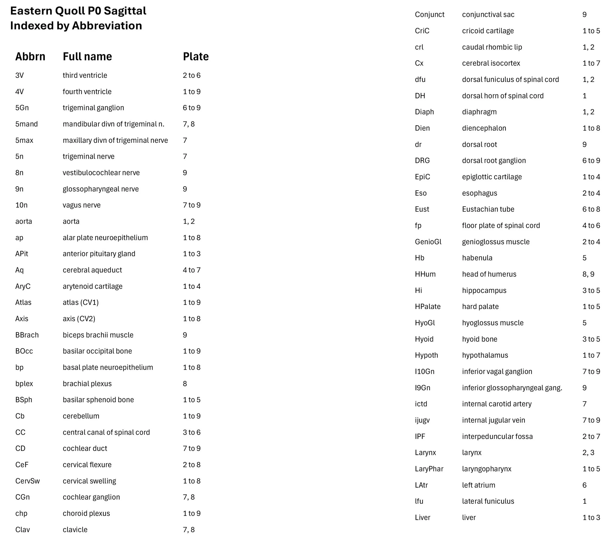
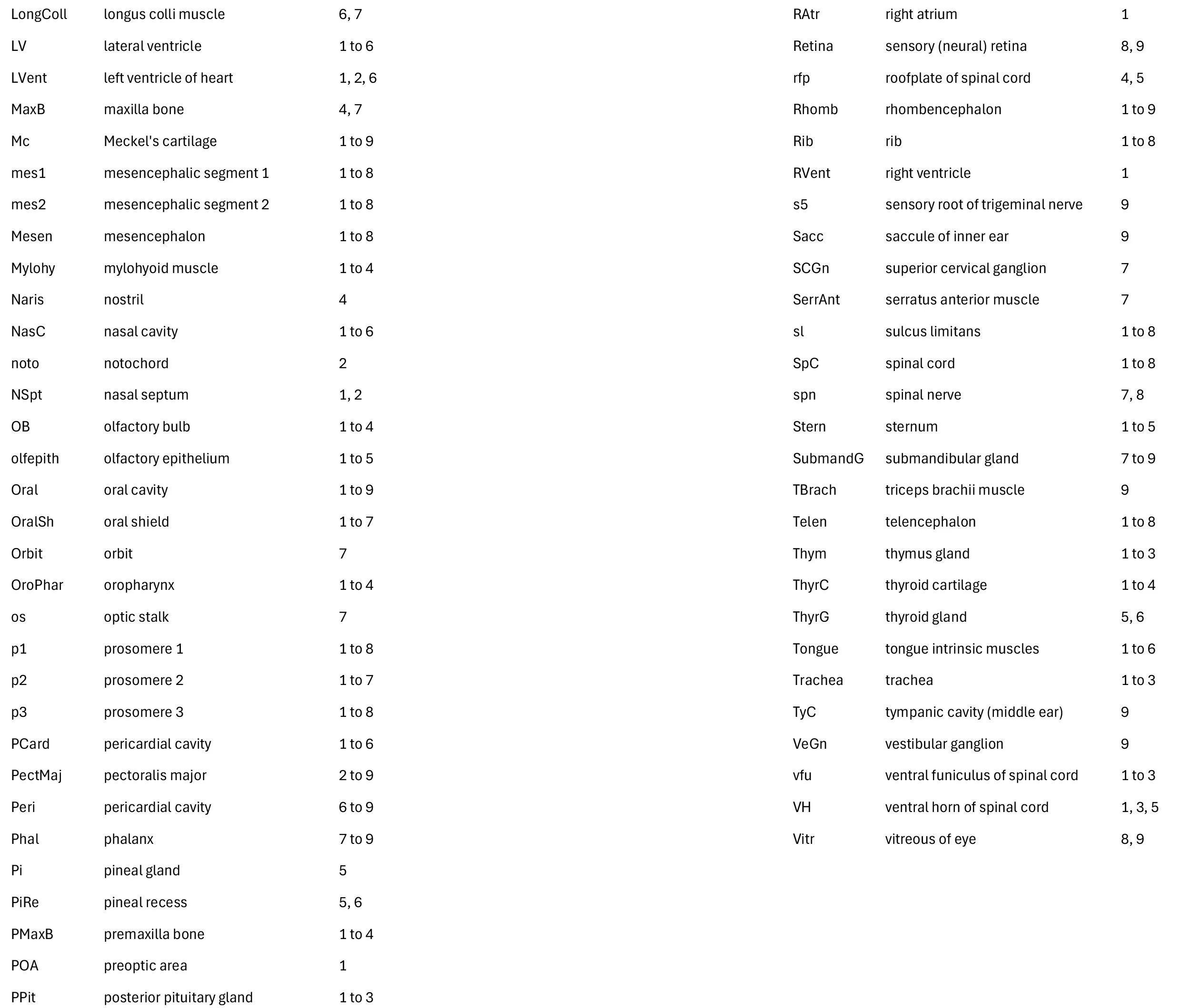
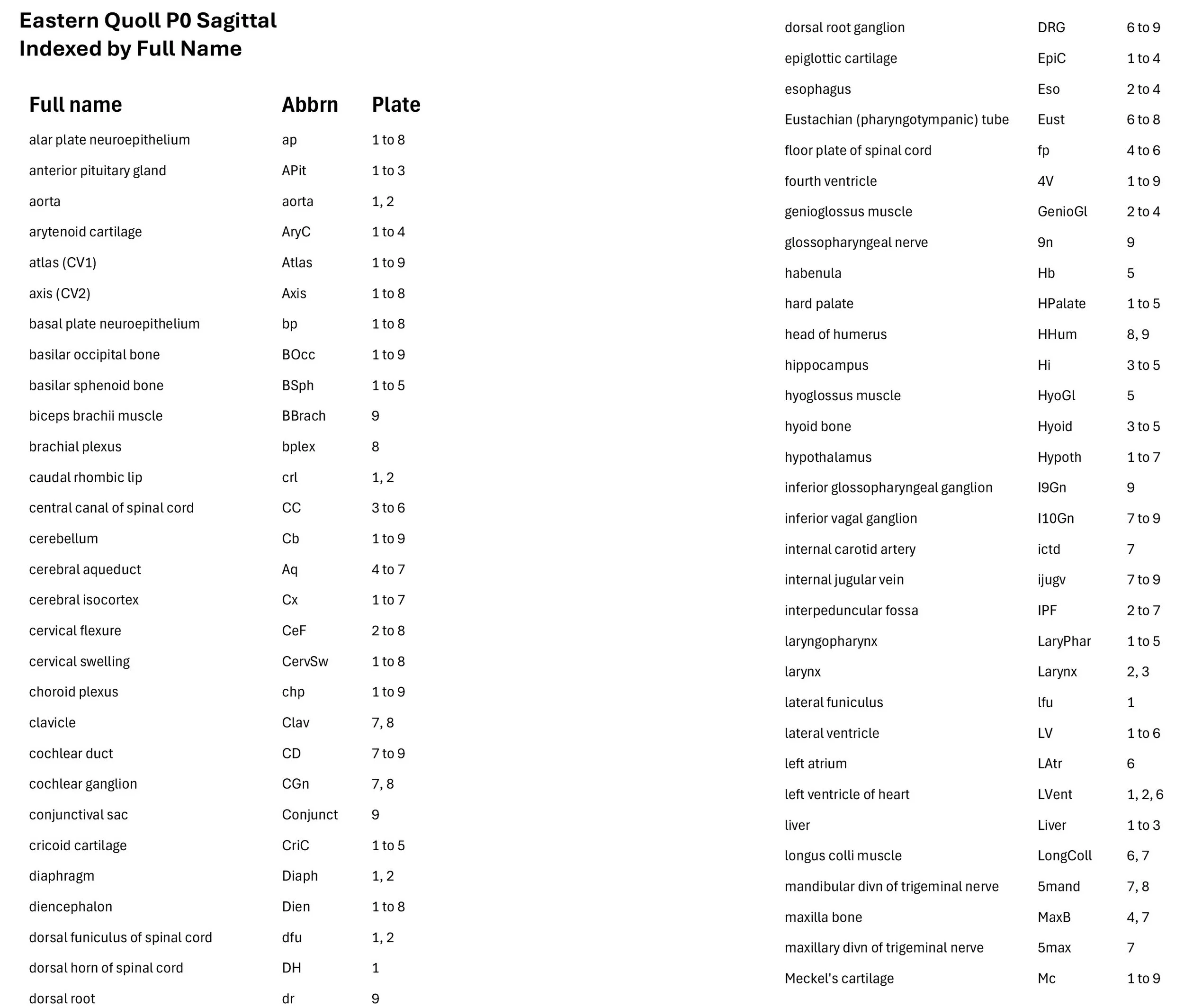
The specimen had been collected in the late 19th century, embedded in paraffin wax and sectioned in a roughly sagittal plane at 10 µm thickness, before being stained with hematoxylin and eosin. The sections at approximately 50 µm intervals were photographed with the aid of a ZeissAxioplan2 fitted with an AxioCamMRc5 camera. All images were calibrated by photographing a scale bar at the same magnification. Images were placed in Adobe Illustrator 2021 and delineated. Developmental regions (i.e. neuroepithelium) destined to give rise to adult structures have been denoted by the adult structure’s name with an asterisk (e.g. Cx* denotes the developmental field of the cerebral isocortex).
Description
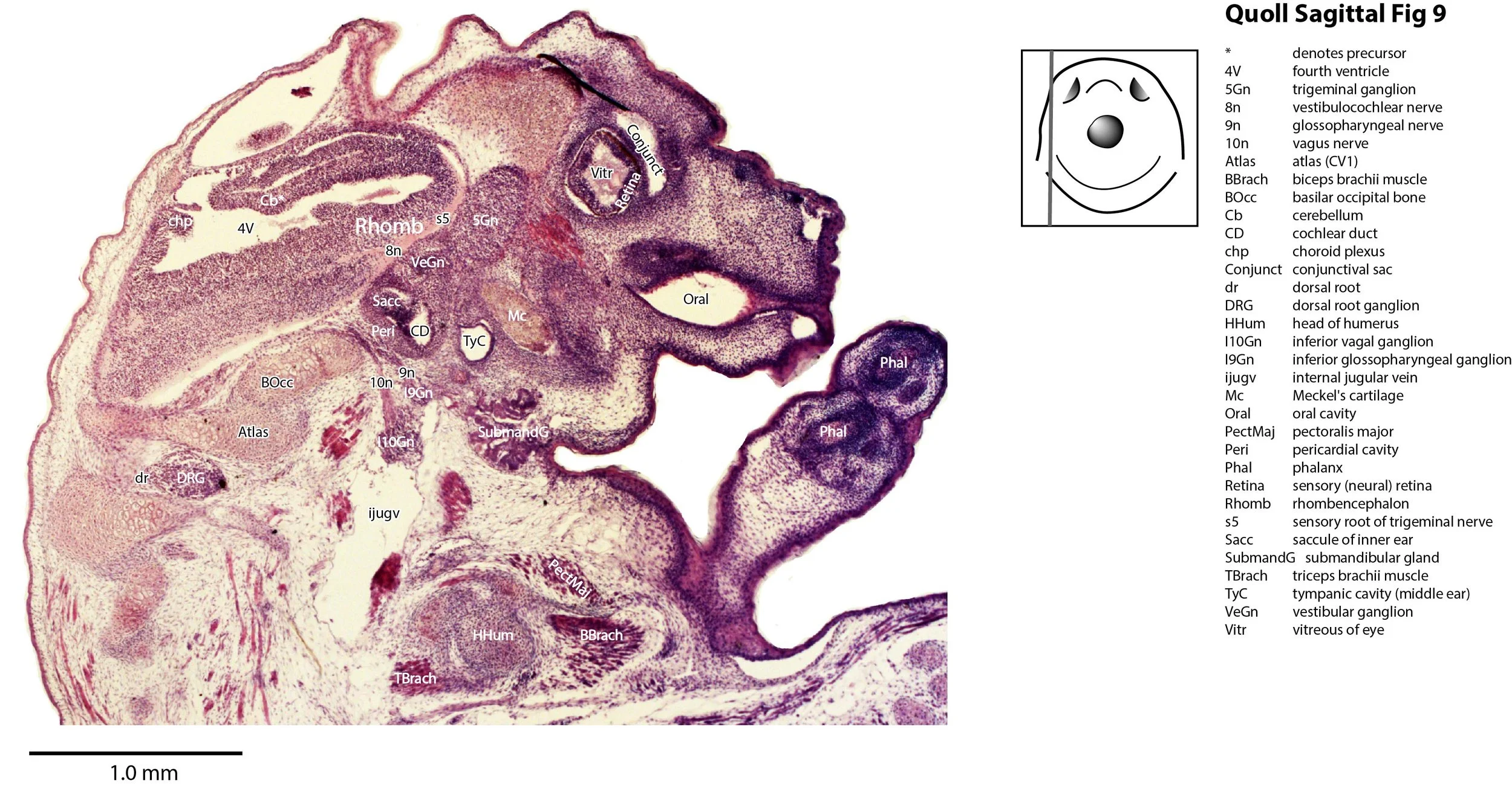
The plane of section is not perfectly sagittal, so not all midline structures appear together in the same section. The tissues of the forebrain (diencephalon and telencephalon) are also poorly preserved, perhaps due to failure of the fixative to fully penetrate these parts of the head. Nevertheless, the key features of brain structure are still visible.
The specimen has a thick oral shield, large oral cavity (presumably for the teat which has been removed) and muscular tongue, but a small nasal cavity. The eyes are rudimentary, being little more than an eye cup of retina. There is a prominent cervical swelling on the chest and upper neck, consisting of loose connective tissue.
The telencephalon is very immature, with a cerebral isocortex and hippocampus that are only neuroepithelium, with no sign of postmitotic neurons. The hypothalamus and diencephalon are also poorly developed, with very few postmitotic neurons present. A pineal recess is visible, but there are no apparent commissures in either the fore- or midbrain. The mesencephalon can be divided into mesencephalic 1 and 2 domains (mes1, mes2), but has only proliferative components dorsally, with some postmitotic neurons ventrally.
The rhombencephalon is the most mature part of the brain, but it is difficult to discern nuclear groups within its postmitotic populations of neurons. Rhombomeric boundaries are also no longer visible. The cerebellum is little more than a ledge of neuroepithelium (rostral rhombic lip) with no post-mitotic populations.
The spinal cord has a thick neuroepithelium, but the ventral horn has emerged to control the forelimb muscles. The dorsal horn is still rudimentary.
Acknowledgements
I would like to thank Dr Peter Giere of the MfN, Berlin Germany, for access to the collection and for all his kind help during the work.