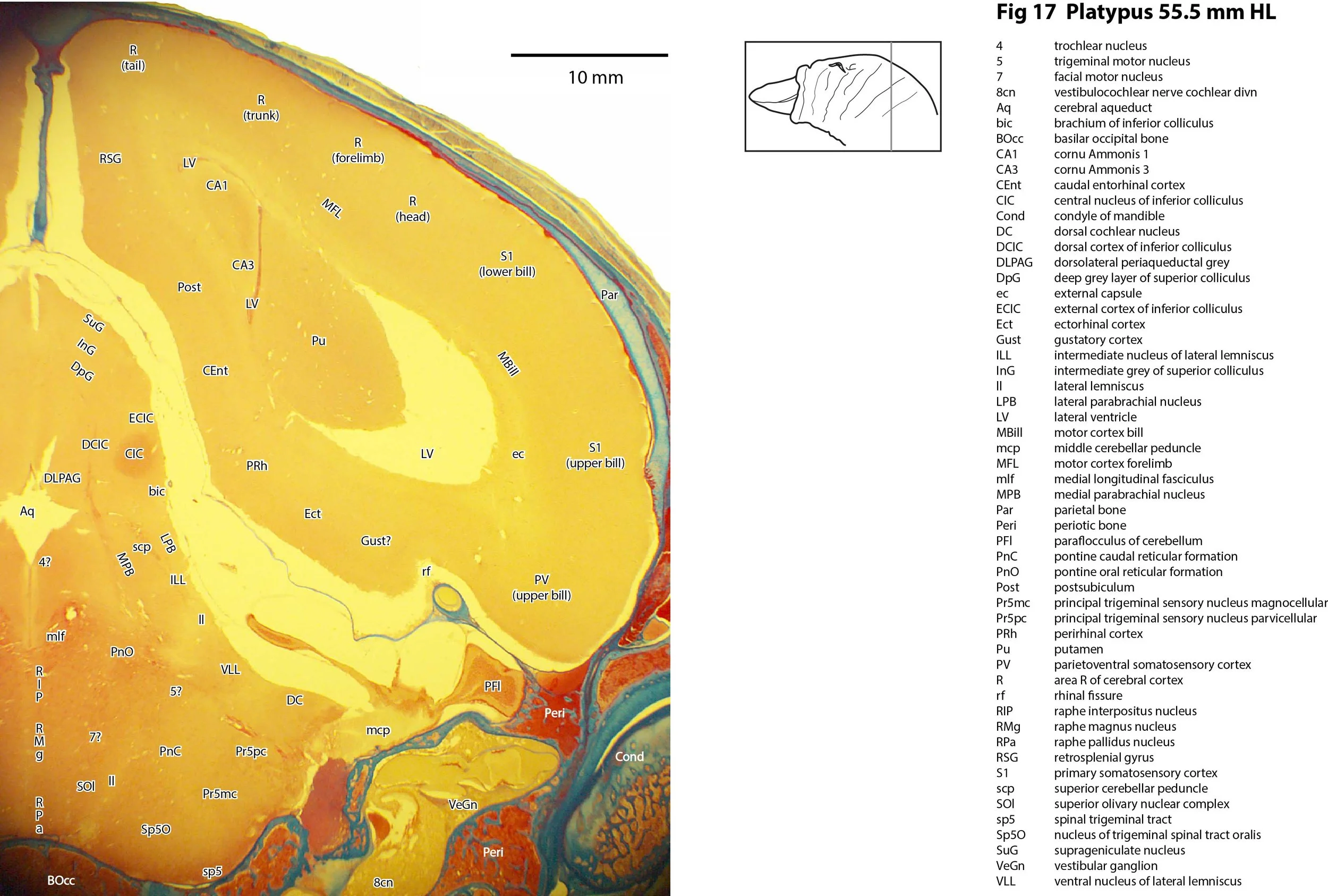
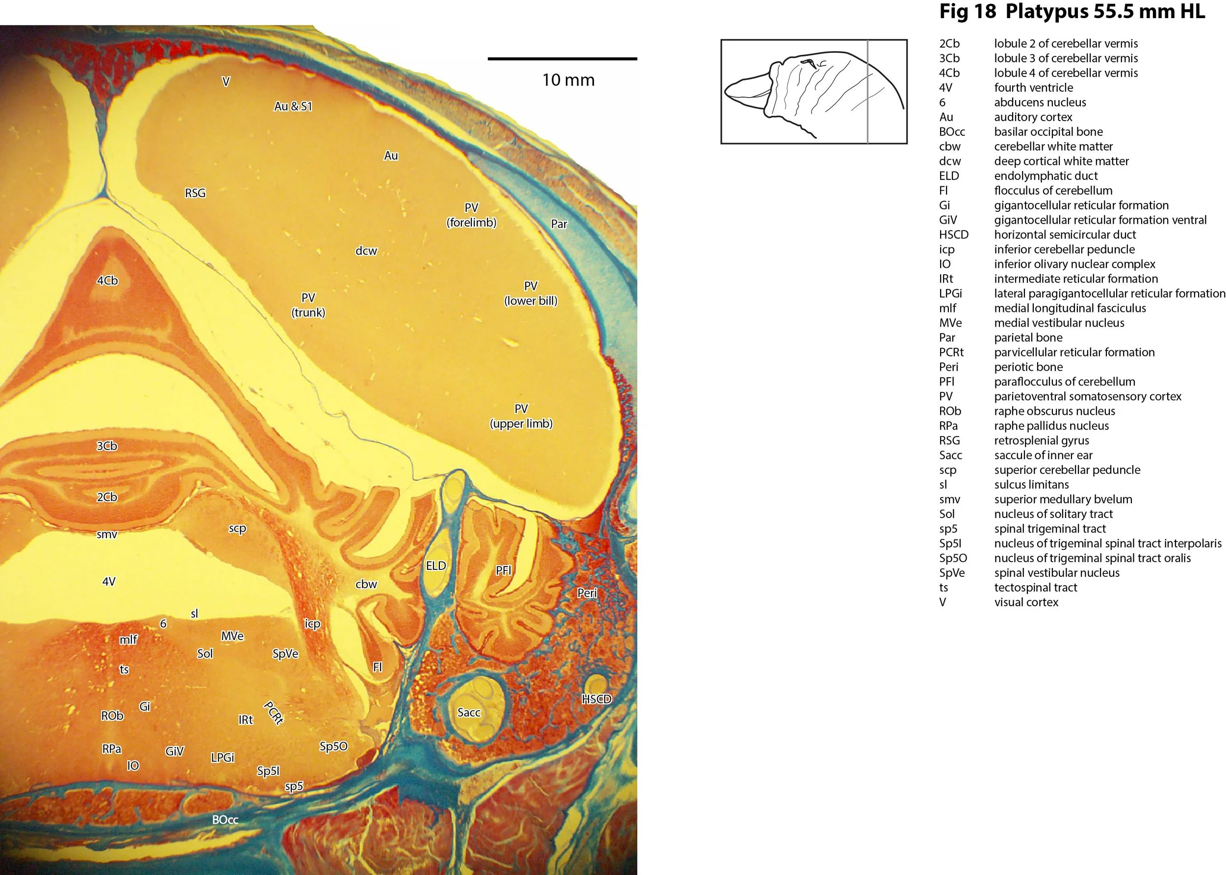
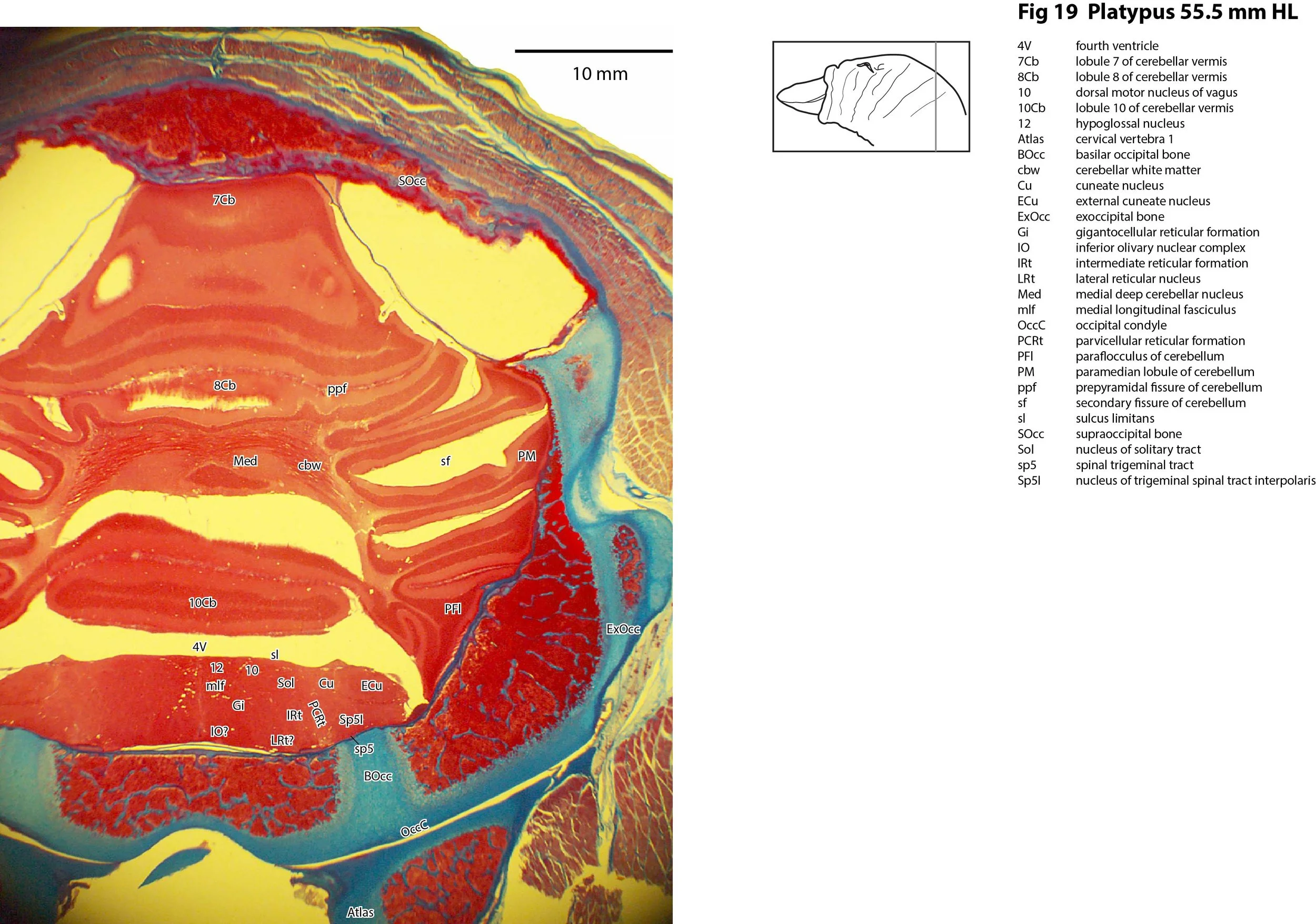
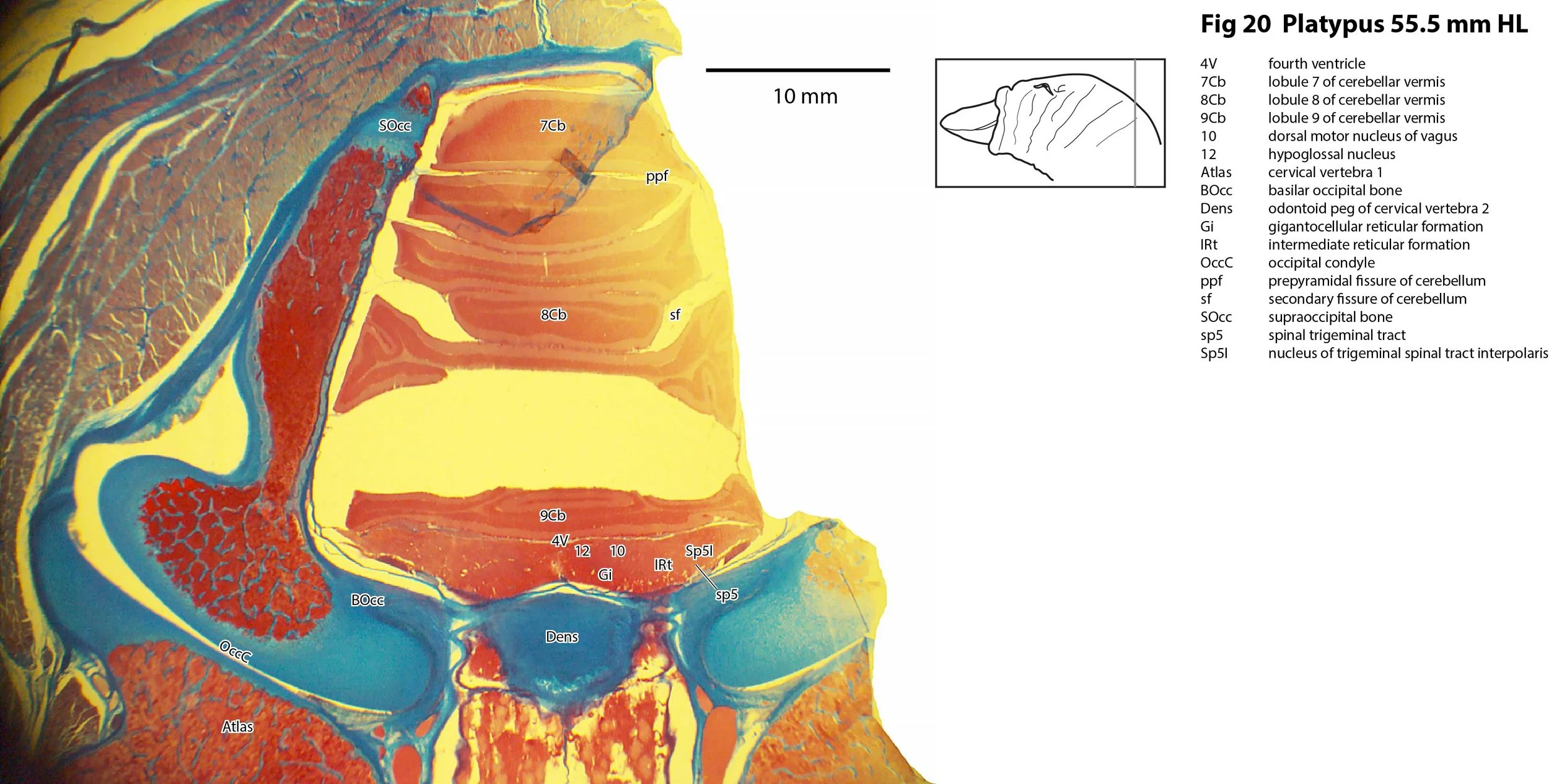
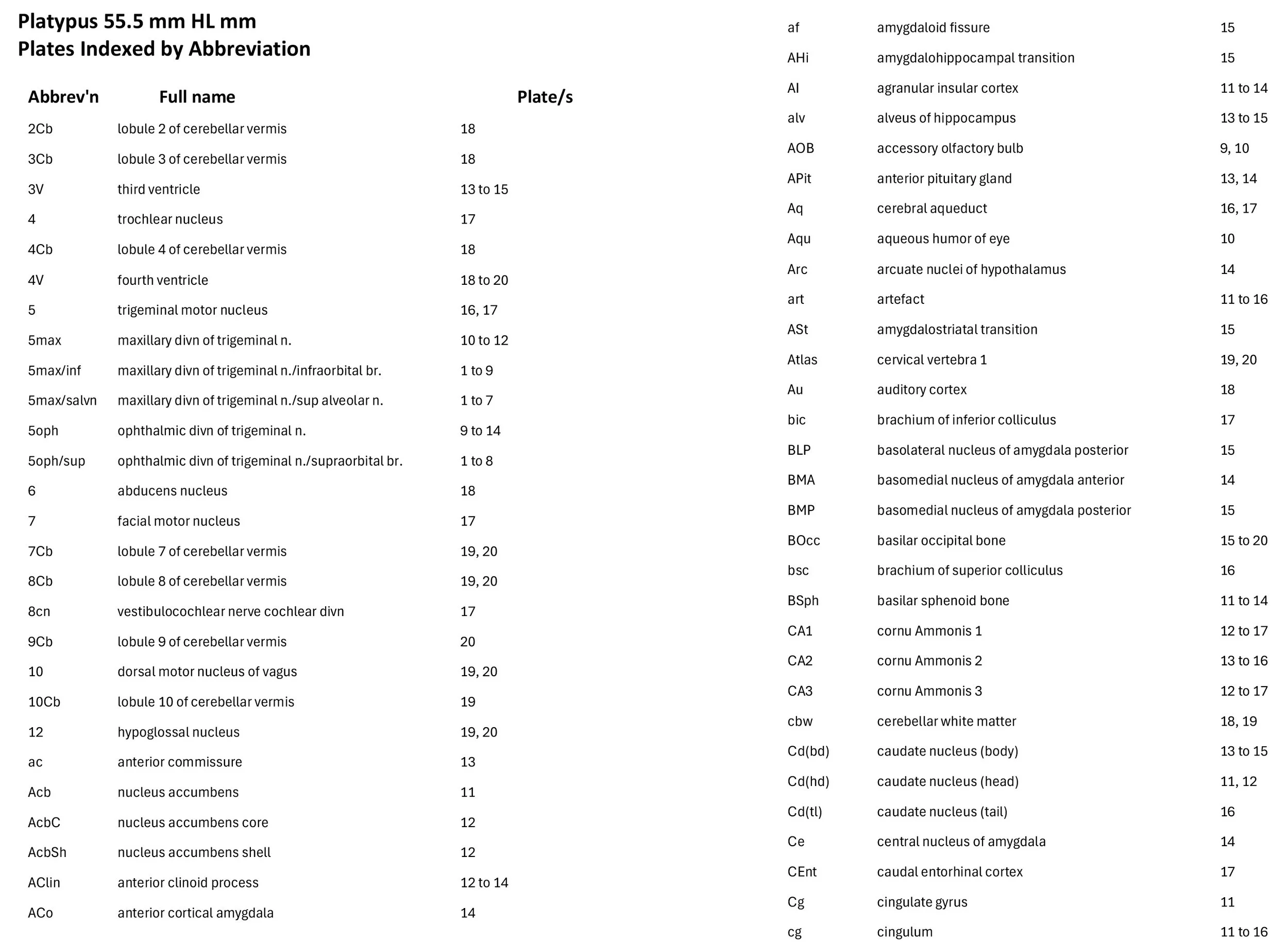
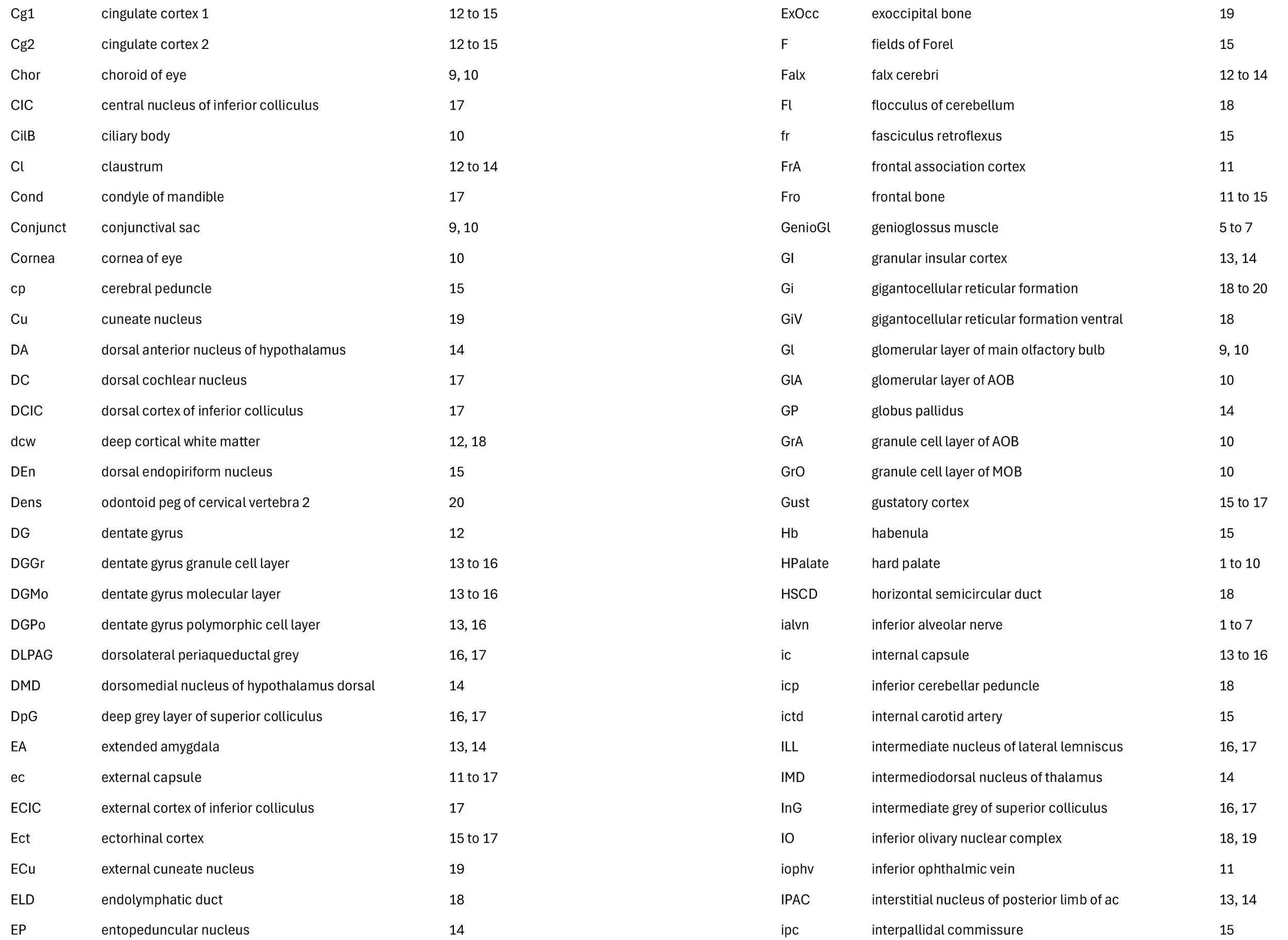
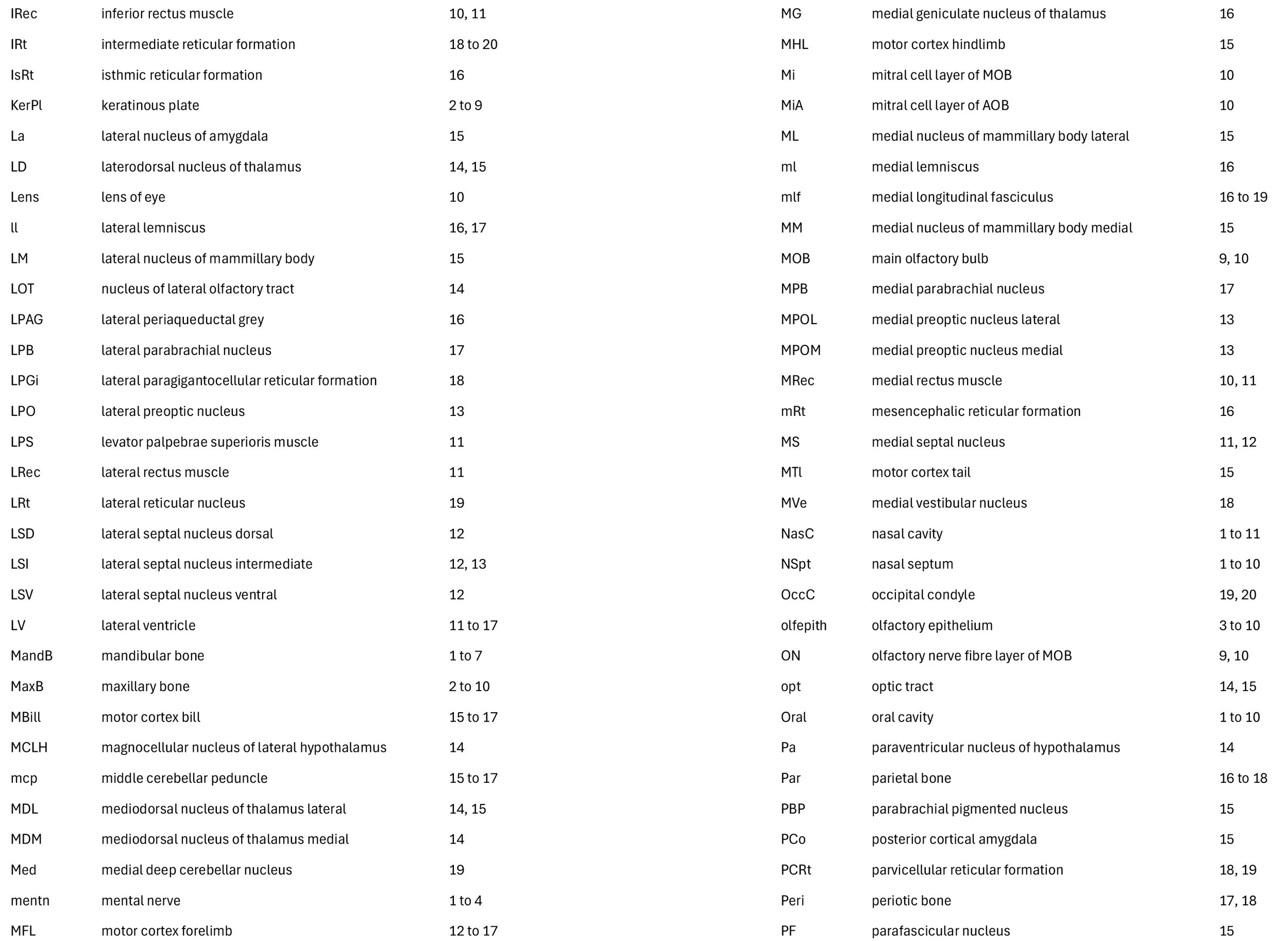
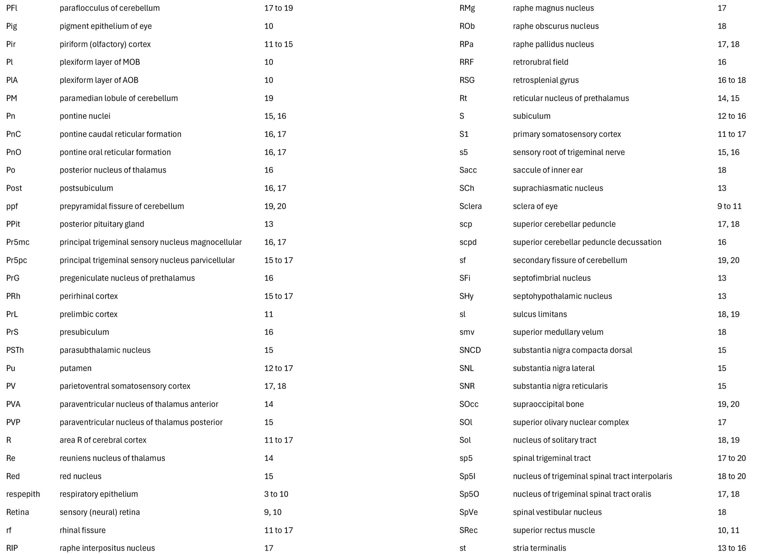
Atlas of the Head of a Juvenile (55.5 mm head length) Platypus
Introduction
Details on platypus biology can be found at the Platypus page of this website. Please see Platypus Brain for topography of the adult cerebral cortex.
Methods
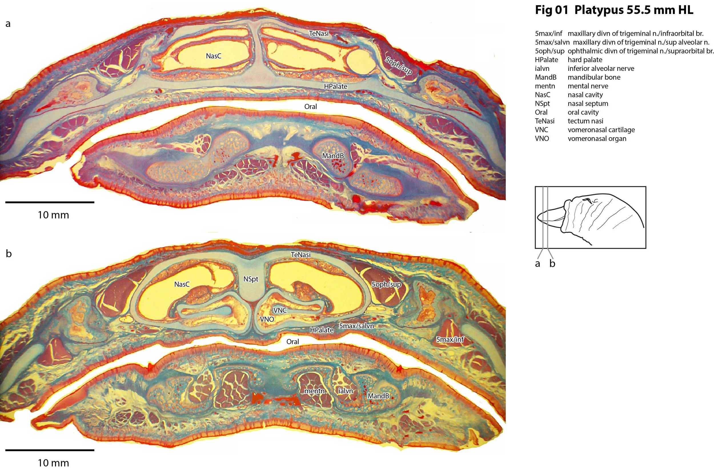
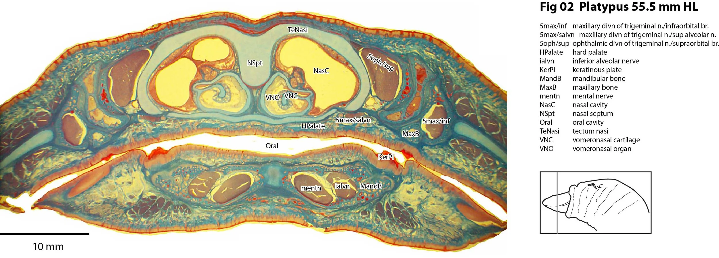
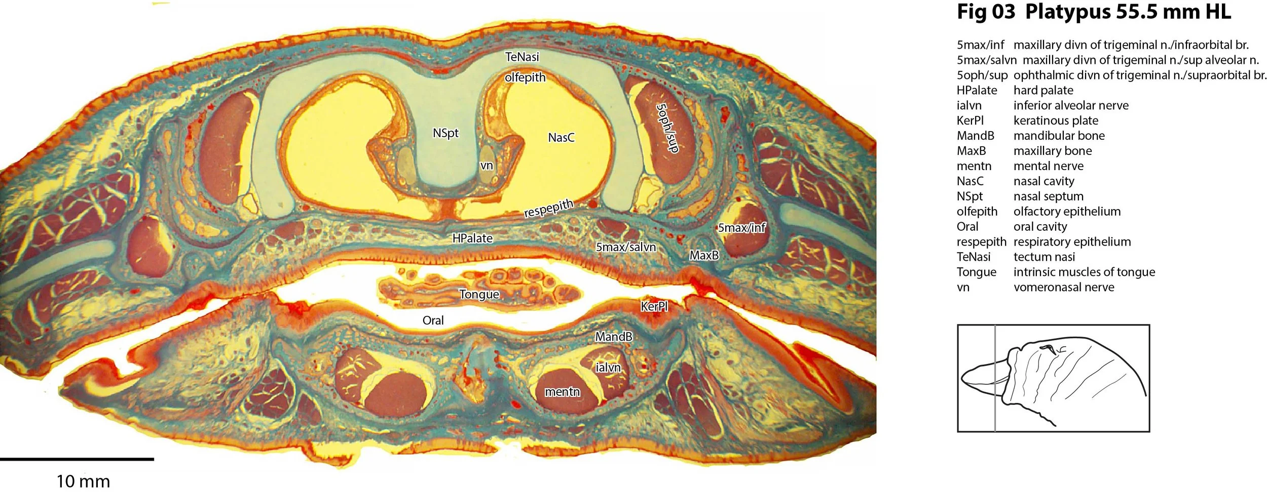
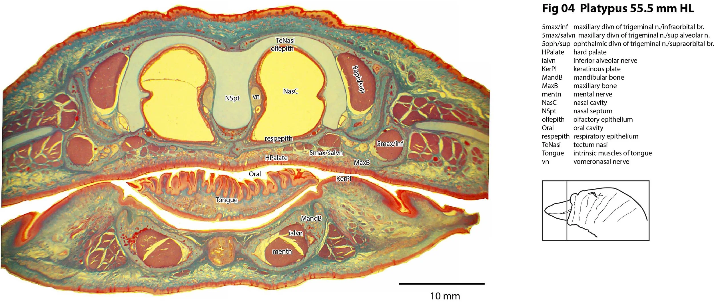
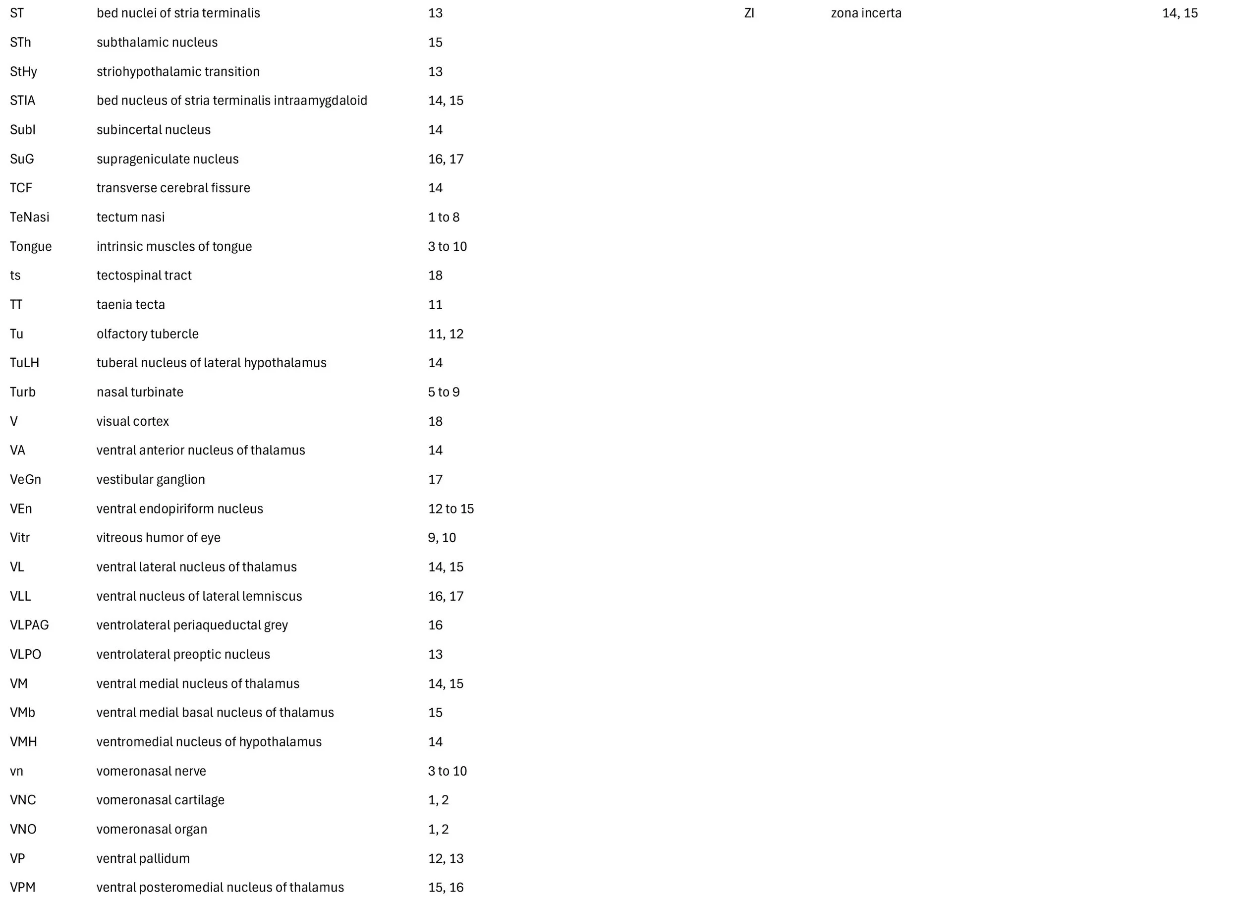
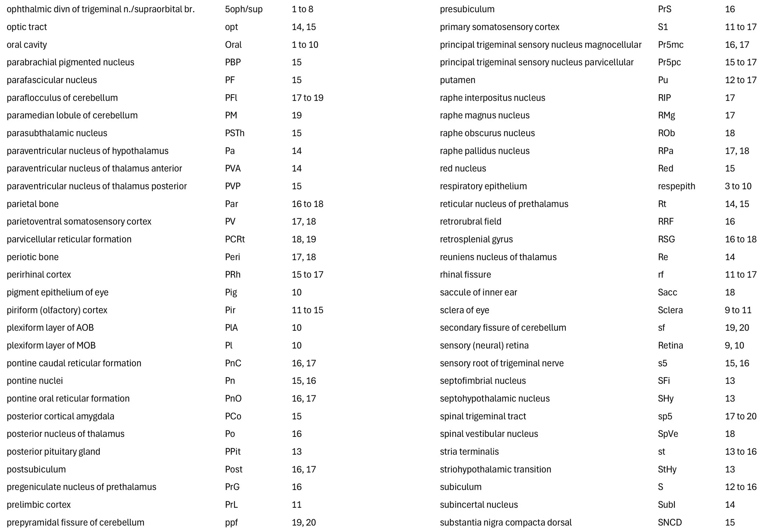
The specimen illustrated here (AMNH201312; head length of 55.5 mm) was kindly made available by Prof. Ulrich Zeller of the Museum für Naturkunde, Berlin (see Zeller, 1988, 1989). The fixation for the specimen is unknown. A head length of 55.5 mm suggests a post-hatching age of approximately 60 days (Ashwell, 2013). The specimen had been decalcified and embedded in Paraplast before being sectioned frontally at a thickness of 35 µm and stained with Azan. The Azan stain is suboptimal for revealing details of brain nuclei and the contrast of grey matter with fibre bundles, but key nuclei are still visible.
General observations
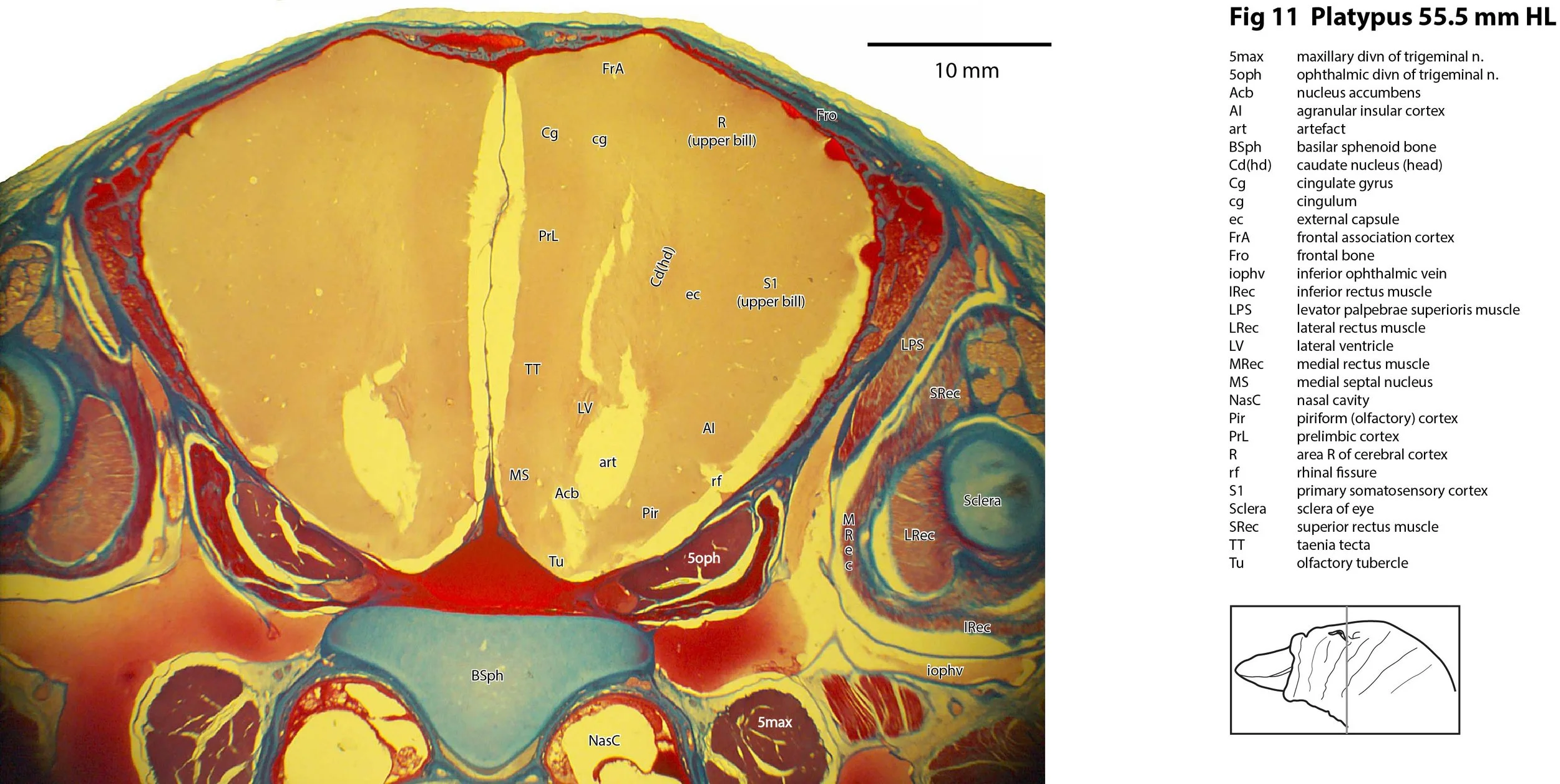
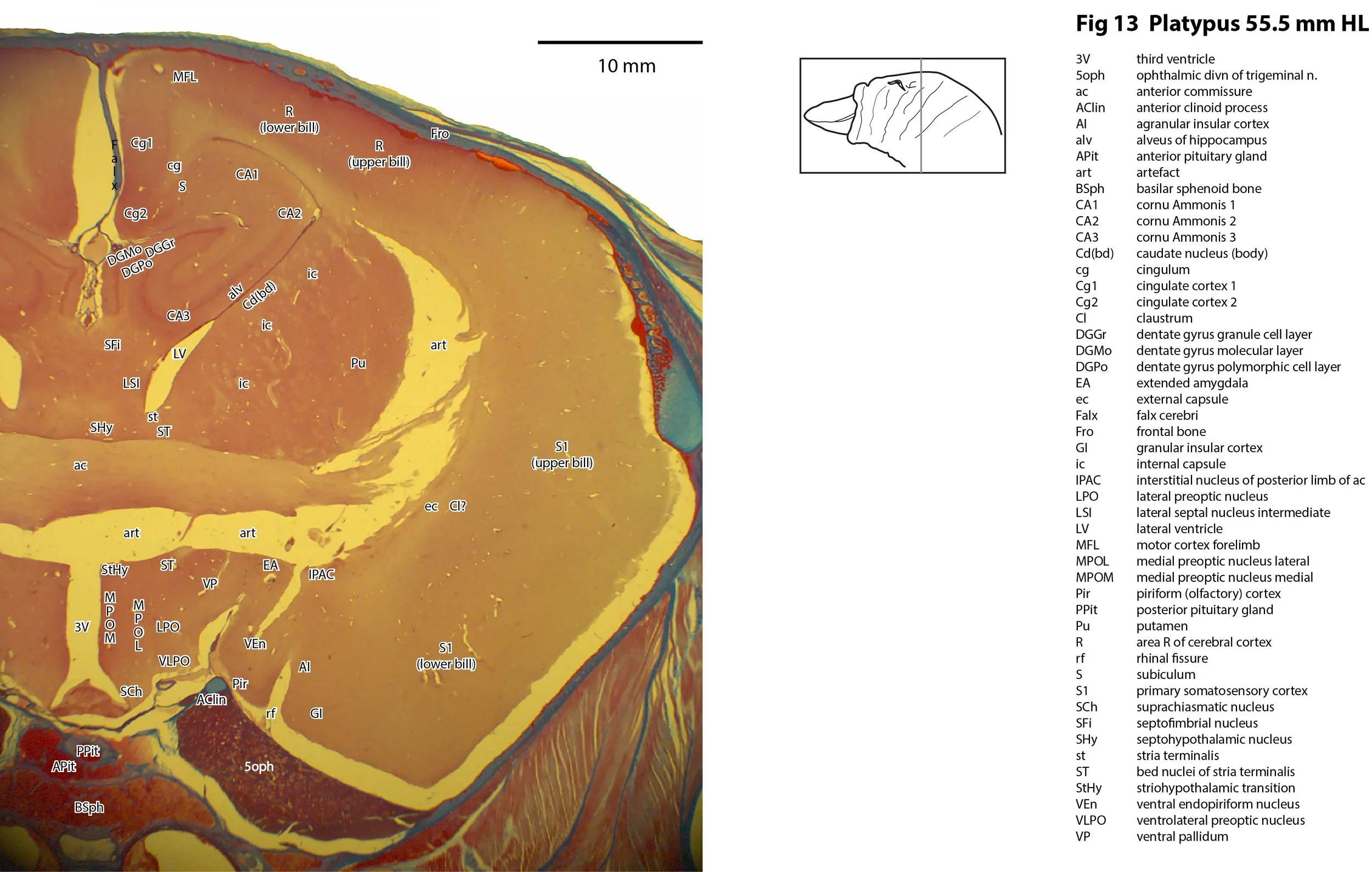
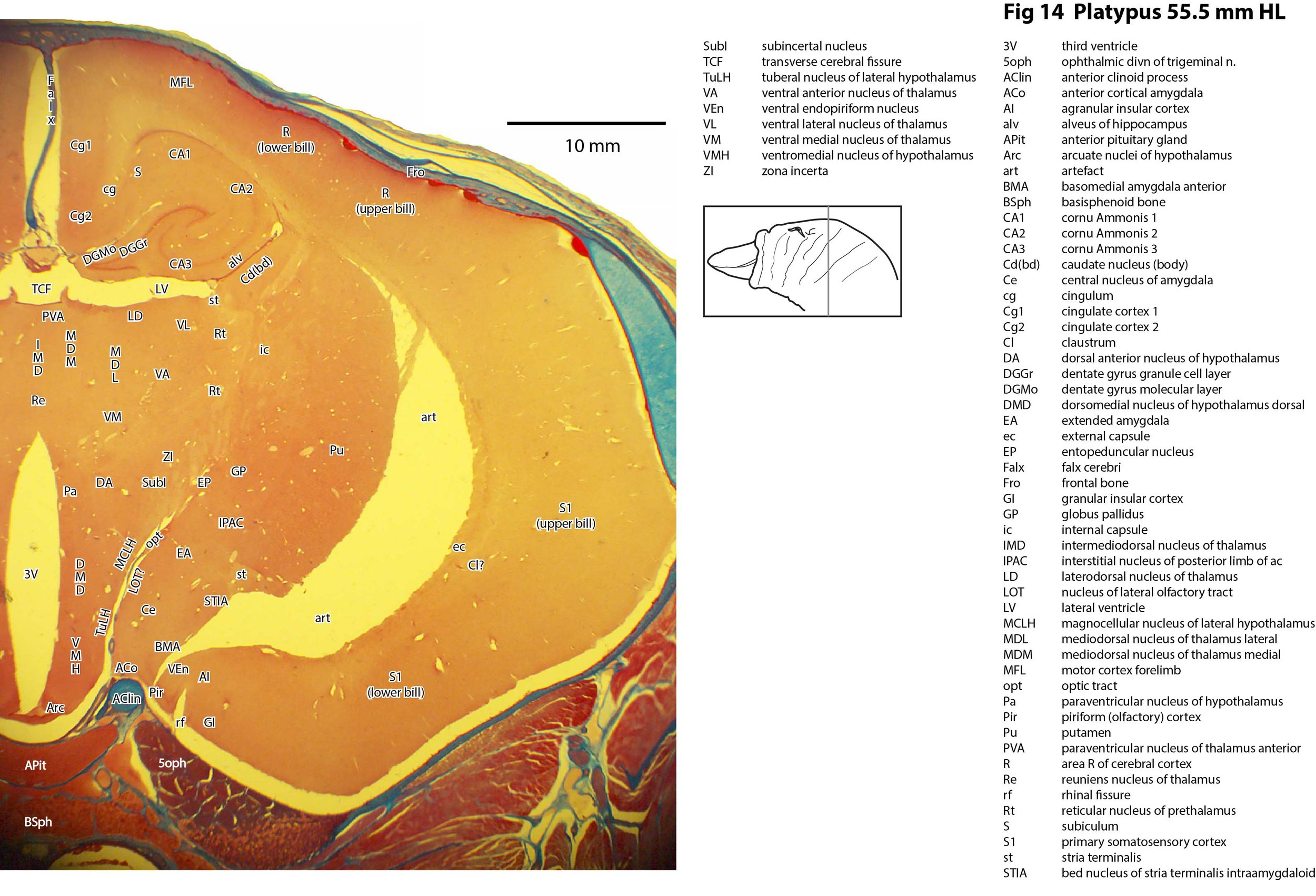
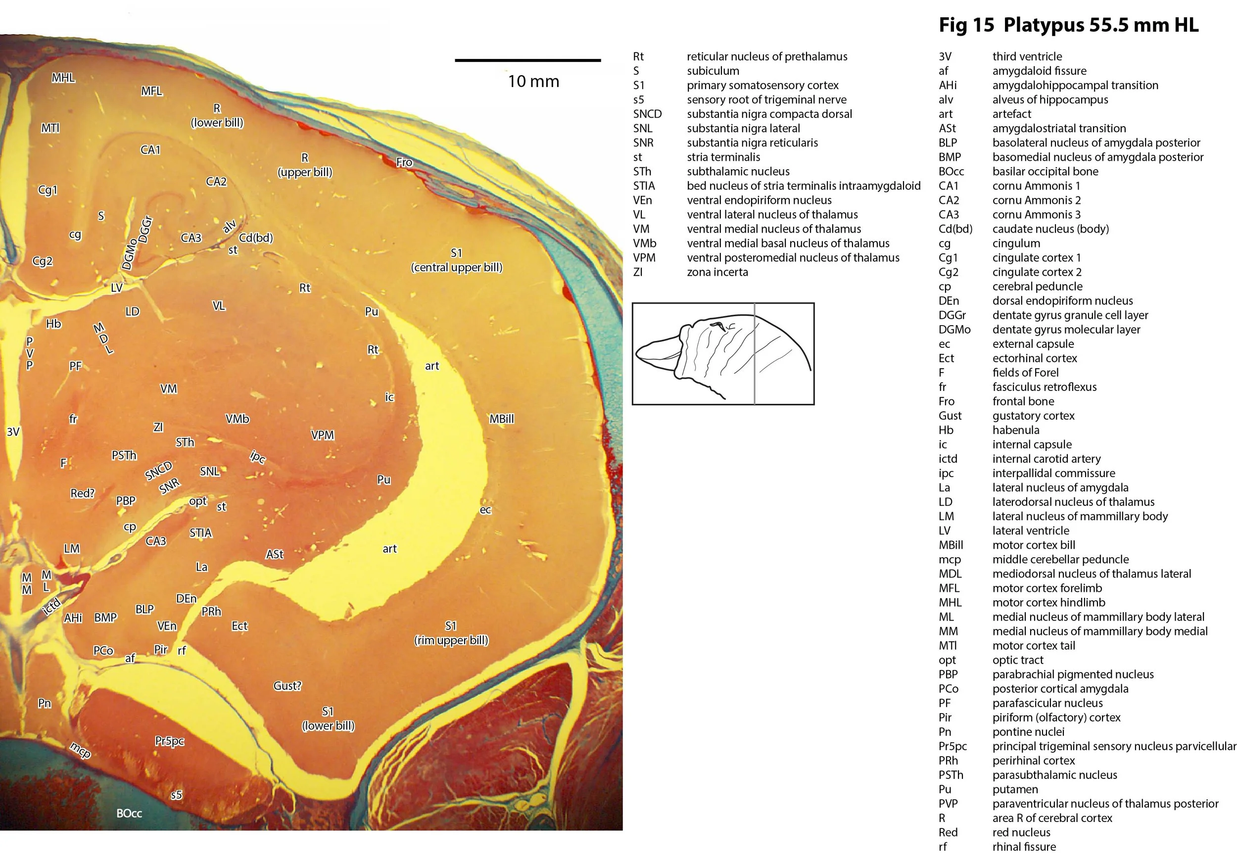
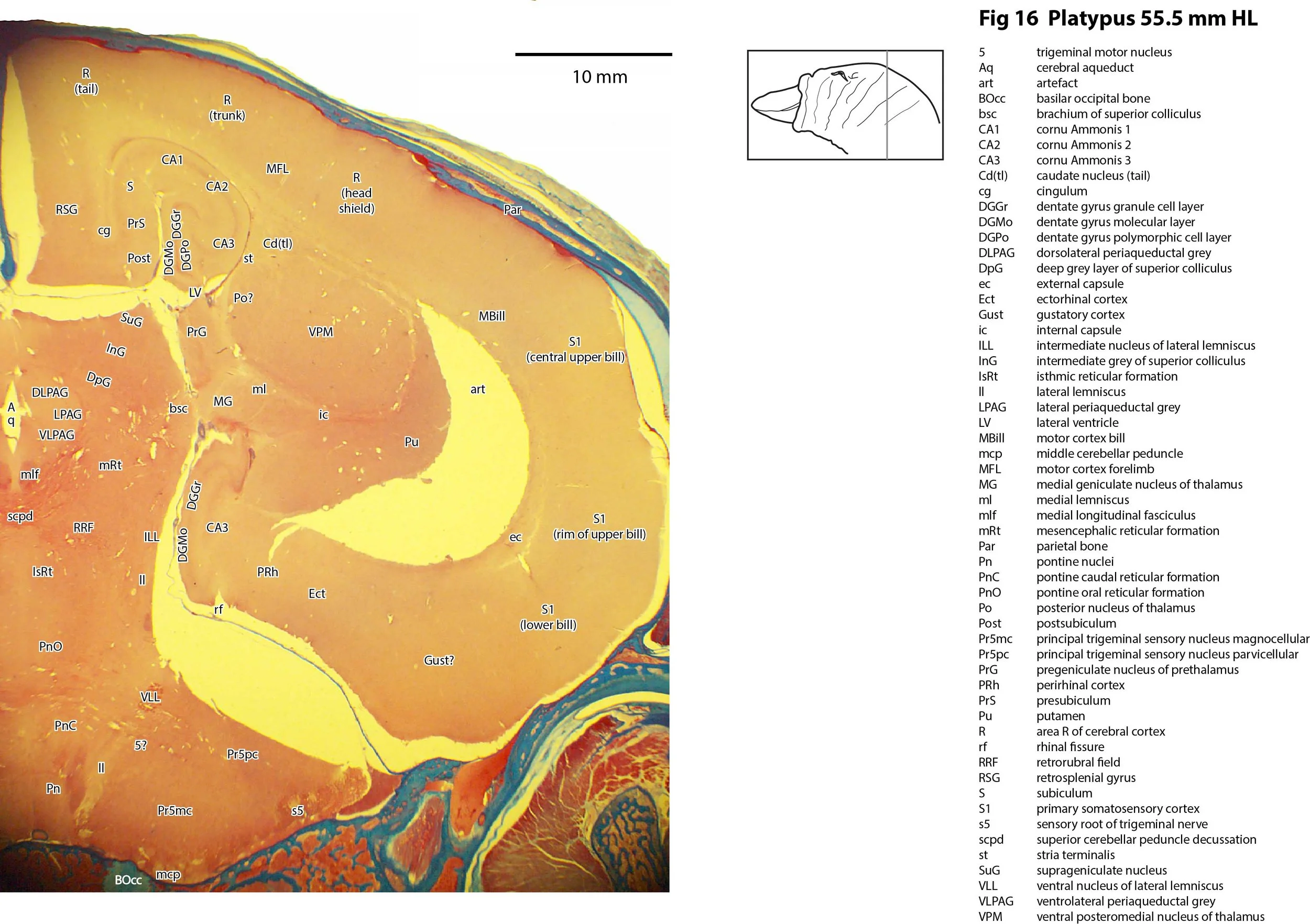
The brain and peripheral nervous system are essentially mature in this juvenile. The stain does not allow all the cellular details of the brain regions to be shown, but all components of the mature nervous system are present. An artefactual tear (art in Plates 11 to 16) is present throughout most of the forebrain.
Trigeminal nerve branches
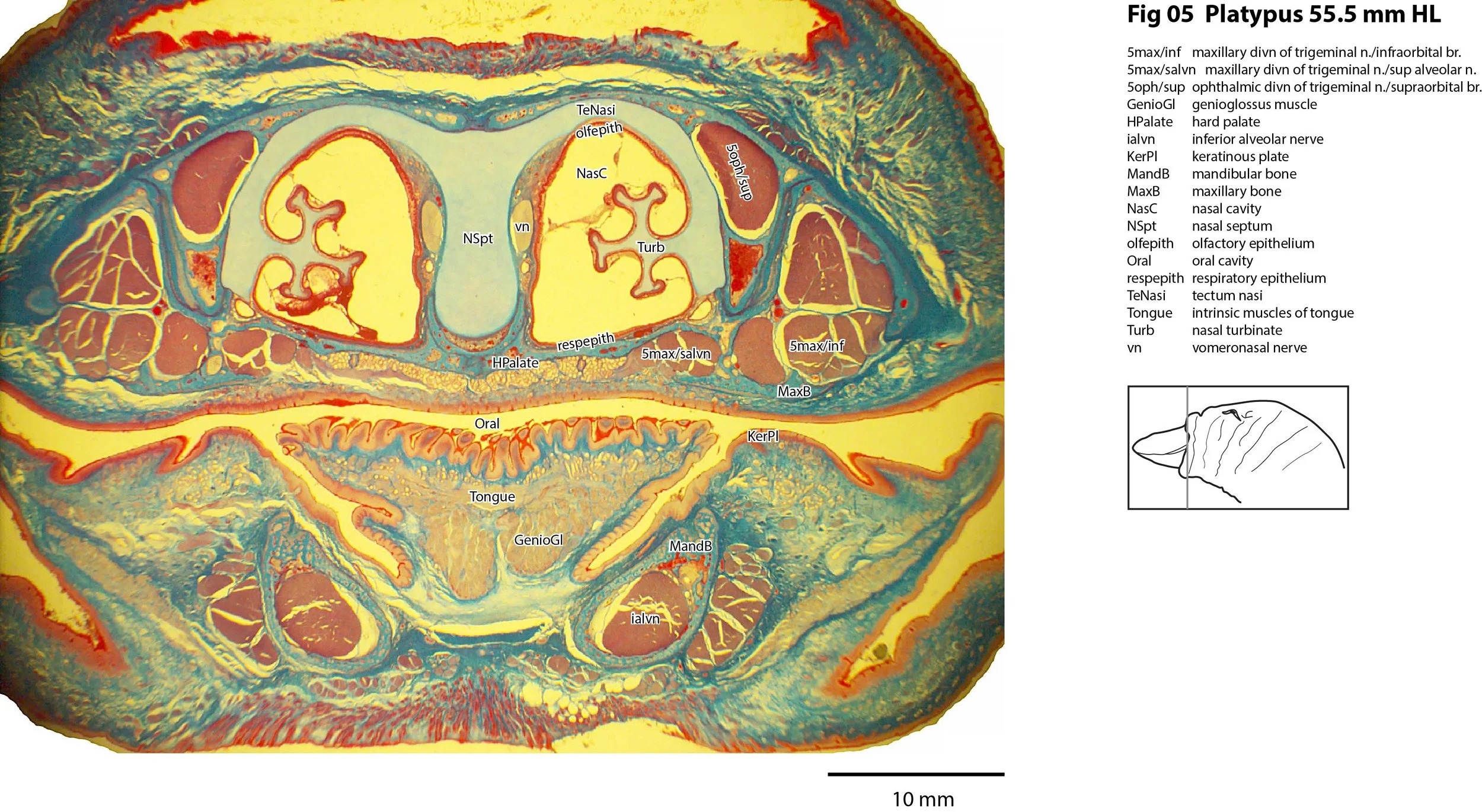
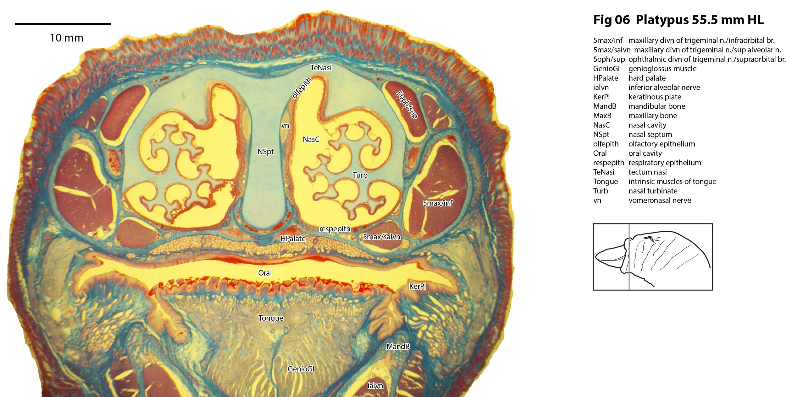
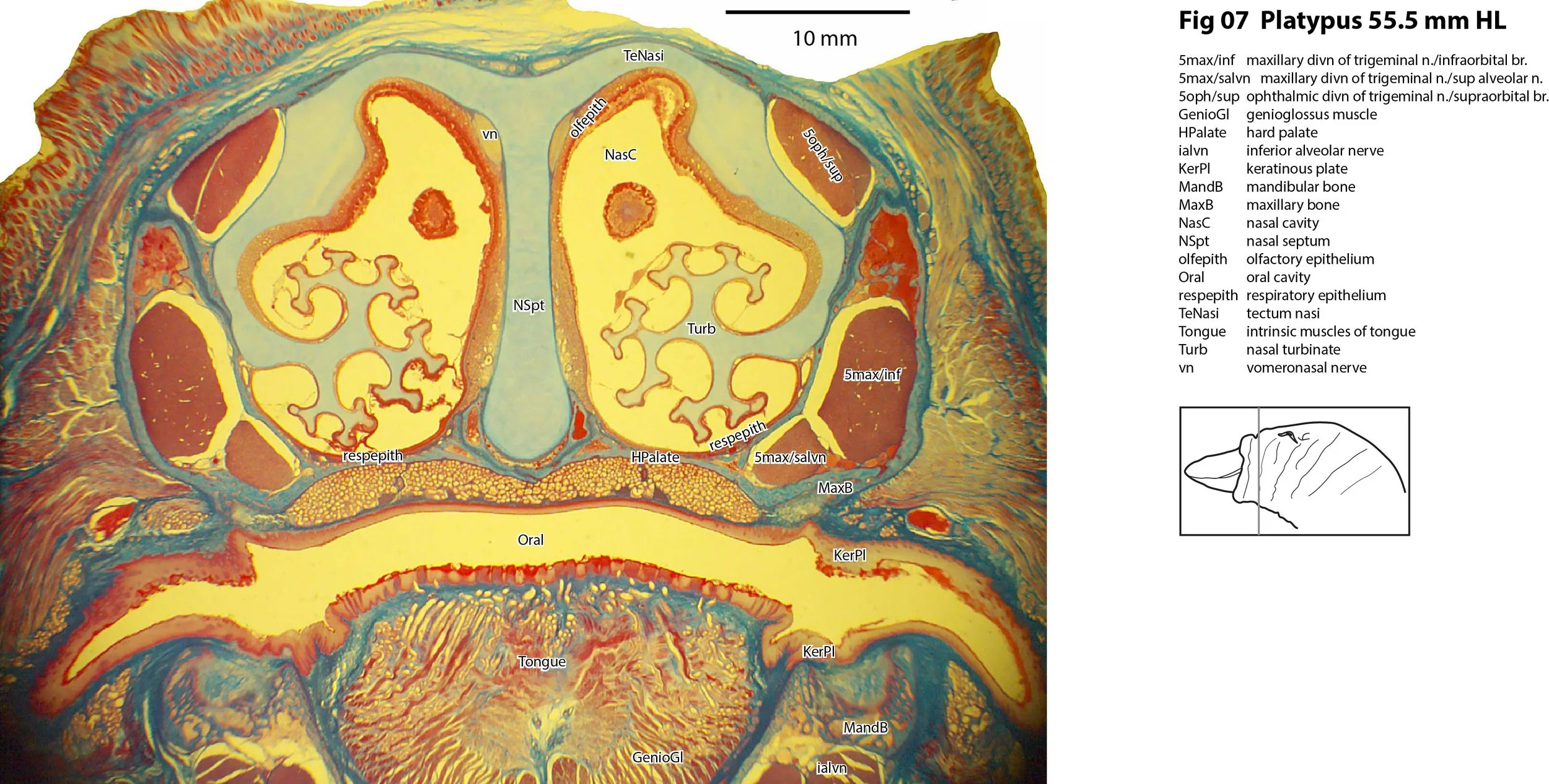
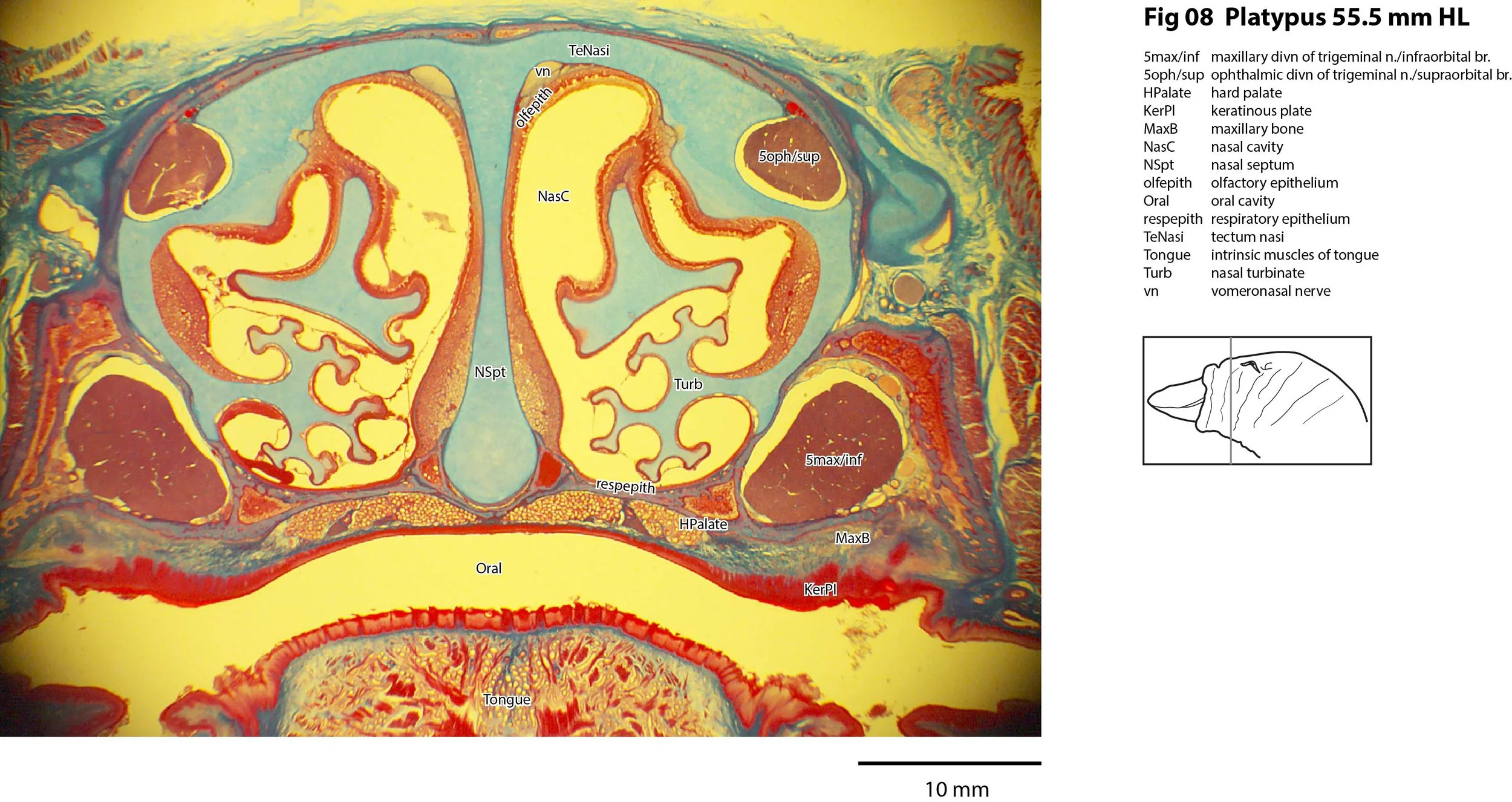
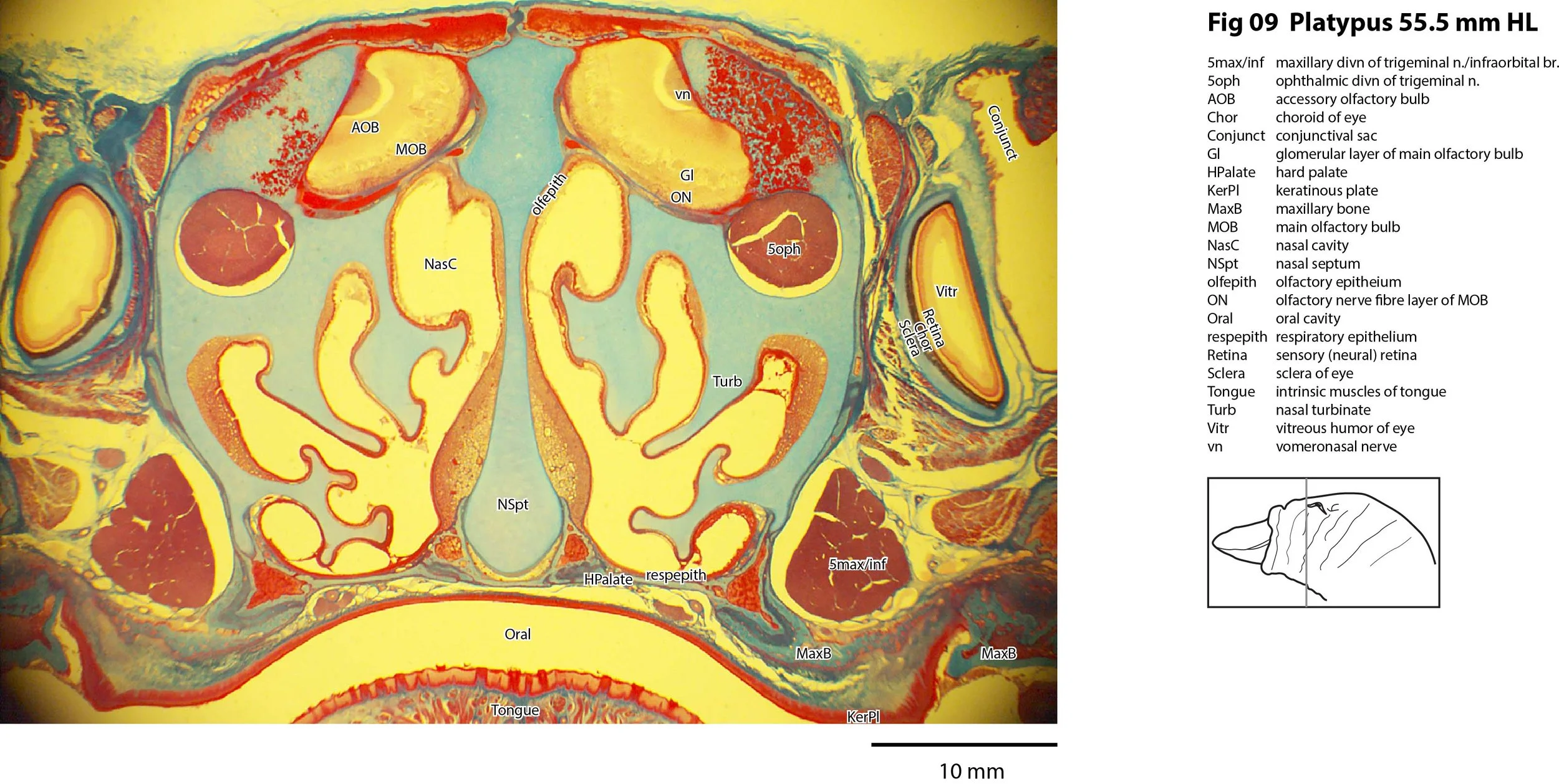
The behaviourally important trigeminal nerve and its divisions (5oph, 5max, 5mand and subdivisions; Plate 9 to 14) are very large. 5oph and 5max provide sensory reception (electro- and mechanosensation) from the greater part of the upper bill. A superior alveolar branch of 5max (Plates 1 to 7) and inferior alveolar (Plates 1 to 7) and mental nerve (Plates 1 to 4) branches of 5mand are present.
Olfactory apparatus
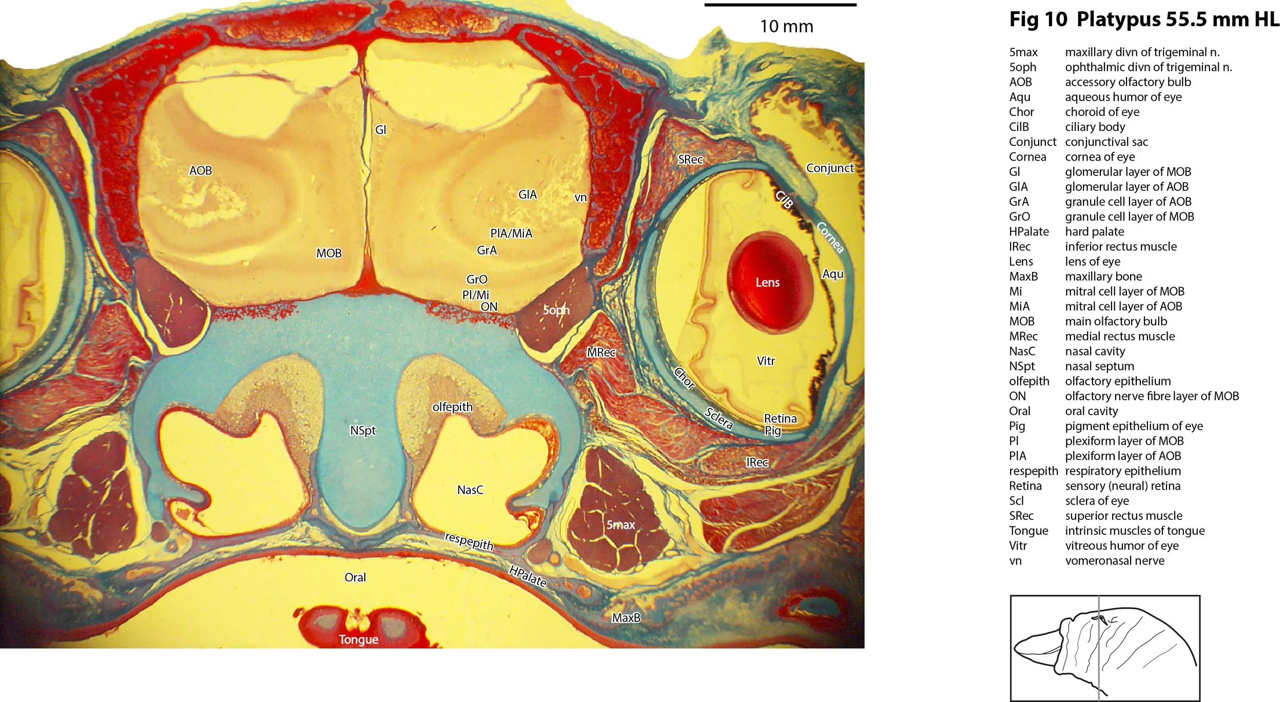
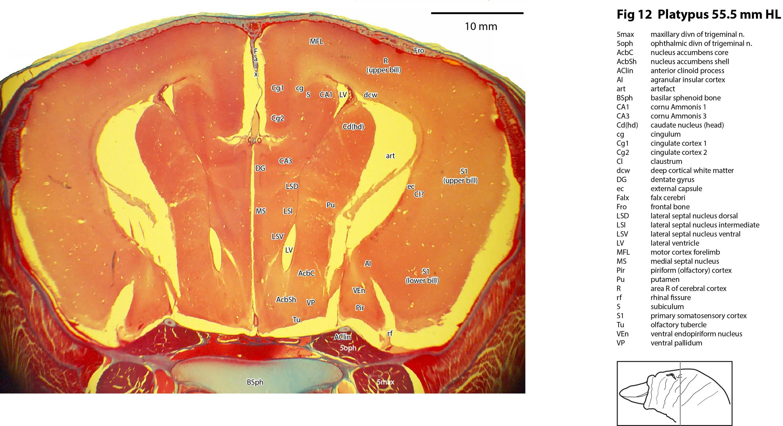
Olfactory epithelium is present in the roof of the nasal cavity (olfepith in Plates 3 to 10). The vomeronasal organ (VNO in Plates 1 and 2) and vomeronasal nerve (vn in Plates 3 to 10) are also visible. The main and accessory olfactory bulbs (MOB, AOB) are mature (see Plates 9 and 10). The piriform (primary olfactory) cortex is mature (Pir in Plates 11 to 15).
Cerebral isocortex (neopallium)
The main functional regions of the cerebral isocortex (S1, R, PV, C, V etc; see Krubitzer et al., 1995) have been indicated, but these are based on topographic position and inherently notional because of the staining methodology.
Hippocampus (archipallium)
The hippocampus (Hi in Plates 12 to 17) is mature and the dentate gyrus can be distinguished (Plates 13 to 16).
Subpallial parts of the telencephalon
Putative nuclei of the amygdala, caudate, putamen and septum have been indicated, but these are necessarily notional because nuclear boundaries are indistinct with the Azan stain.
(Dorsal) thalamus
The dorsal thalamus (prosomere 2 of diencephalon) doesn’t show clear internal subdivisions so the labelling of constituent nuclei is necessarily notional and based on topography.
Cerebellum
The cerebellum is mature and the external granular (germinal) layer that produces the microneurons of the cerebellar cortex has involuted. Major components of the cerebellar hemispheres have been indicated.
Rhombencephalon
The Azan stain allows only an indistinct delineation of medullary nuclei, but the major cranial nerve nuclei can be distinguished. The most striking feature of the brainstem is the large size of the trigeminal sensory column (Pr5pc, Pr5mc, Sp5O, Sp5I; see Plates 15 to 20), particularly the most rostral elements close to the trigeminal nerve (5n; see Plates 15 to 17) entry. See Ashwell et al. (2006) for details of adult trigeminal nuclei in this species.
Acknowledgements
Acknowledgement is given to the American Museum of Natural History who provided this specimen for Prof. Zeller’s work. I would like to thank Prof Ulrich Zeller and Dr Peter Giere of the MfN, Berlin Germany, for access to the MfN collection and for all their help during the work.
References
Ashwell KW (2013) Embryology and post-hatching development of the monotremes. In KWS Ashwell (Ed.), Neurobiology of Monotremes: Brain Evolution in Our Distant Mammalian Cousins (1st ed., pp. 31-46). CSIRO.
Ashwell KWS, Hardman CD, Paxinos G (2006) Cyto- and chemoarchitecture of the sensory trigeminal nuclei of the echidna, platypus and rat. Journal of Chemical Neuroanatomy31, 81-107.
Krubitzer L, Manger P, Pettigrew J, Calford M (1995) Organization of somatosensory cortex in monotremes. In search of the prototypical plan. Journal of Comparative Neurology351, 261–306.
Zeller U (1988) The lamina cribrosa of Ornithorhynchus (Monotremata, Mammalia) Anatomy and Embryology178, 513–519.
Zeller U (1989) Die Entwicklung und Morphologie des Schadels von Ornithorhynchusanatinus: (Mammalia, Prototheria, Monotremata). Abhandlungen der Senckenbergischen Naturforschenden Gesellschaft545, 1–188. Verlag Waldemar Kramer, Frankfurt.